Consequence of lithium therapy is the most likely diagnosis
Ashley Davidoff MD
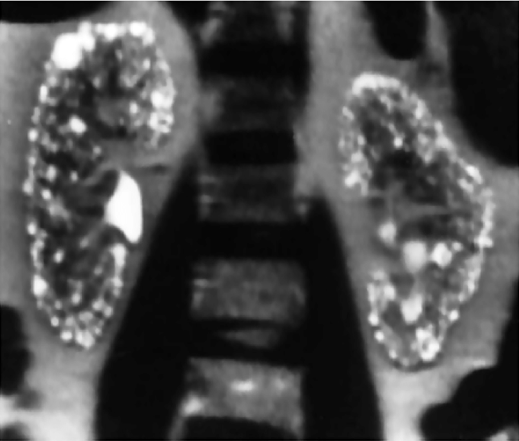
018 72F Asian renal capsular calcification
Consequence of lithium therapy is the most likely diagnosis
Ashley Davidoff MD
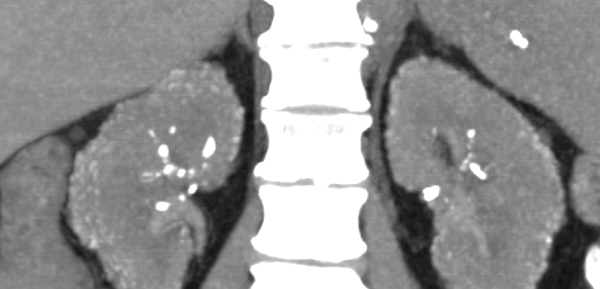
020 72F Asian renal capsular calcification
Consequence of lithium therapy is the most likely diagnosis
Ashley Davidoff MD
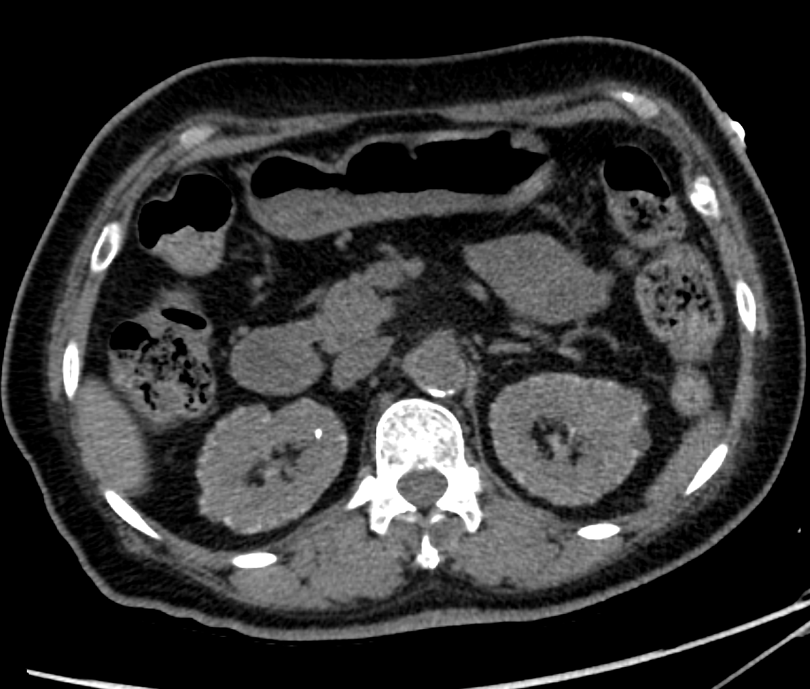
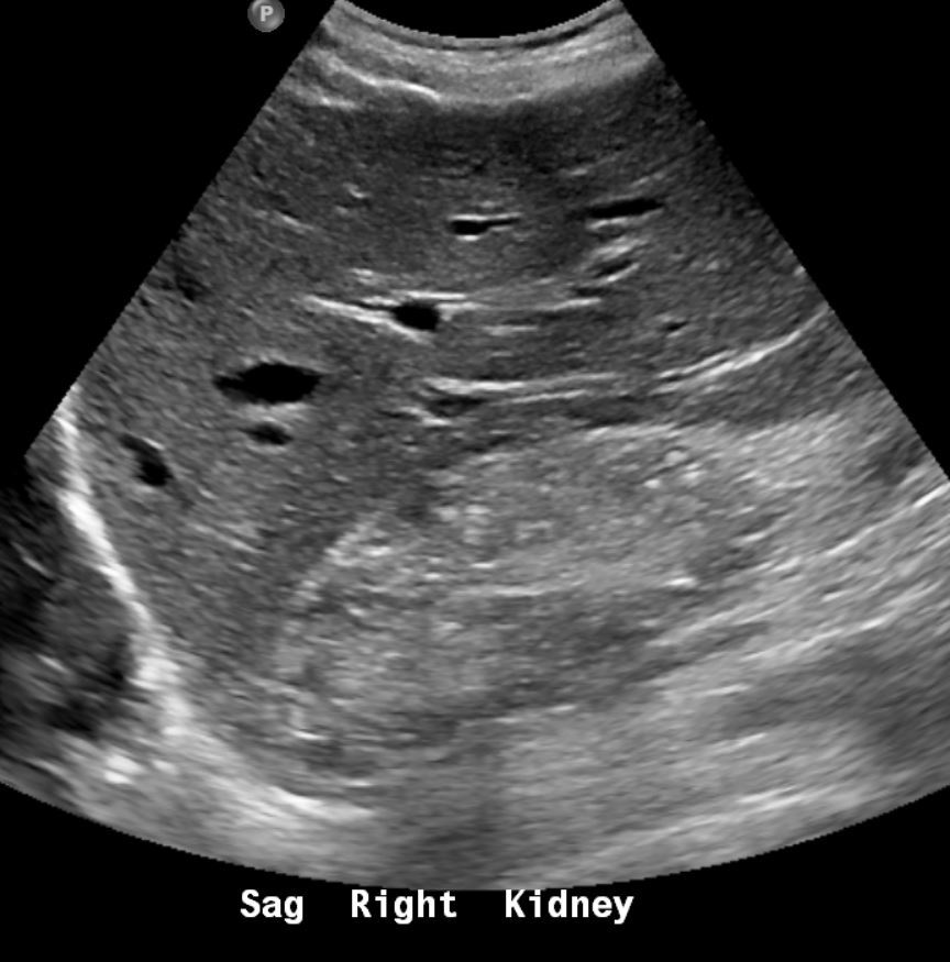
019 72F Asian renal capsular calcification
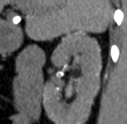
A Second Case
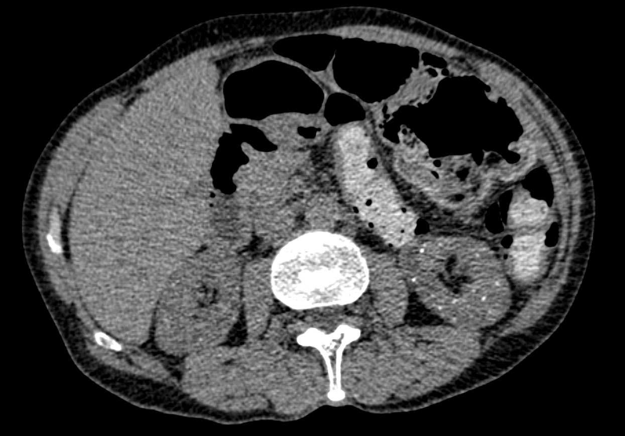
CT shows numerous punctate calcifications in the cortex
and medulla bilaterally. Given history of lithium usage, CT appearance can be
seen with lithium-induced renal disease.
Ashley Davidoff The CommonVein.net
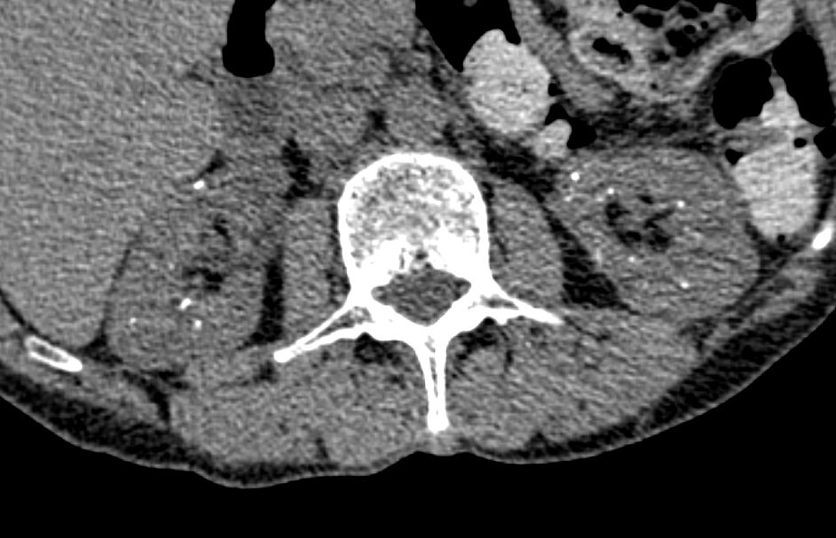
CT shows numerous punctate calcifications in the cortex
and medulla bilaterally. Given history of lithium usage, CT appearance can be
seen with lithium-induced renal disease.
Ashley Davidoff The CommonVein.net
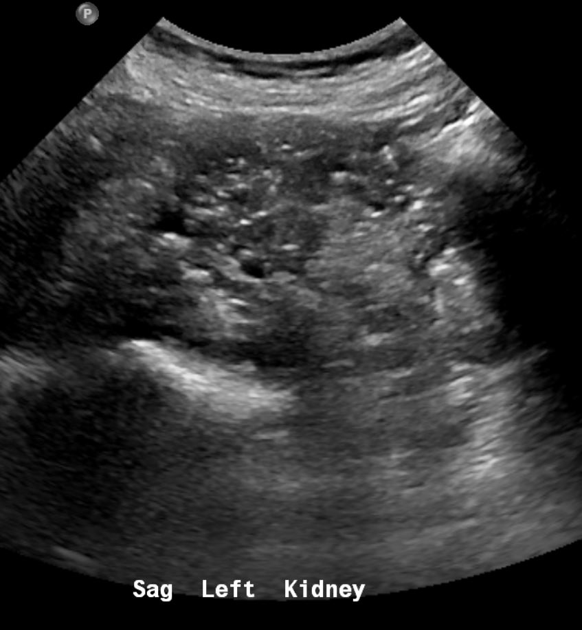
and medulla. Ct showed multiple punctate calcifications Given history of lithium usage, sonographic appearance can be
seen with lithium-induced renal disease.
Ashley Davidoff The CommonVein.net
Lithium used in the treatment of bipolar disorder.
-
- renal toxicity
- impaired urinary concentrating ability
- natriuresis,
- renal tubular acidosis,
- tubulointerstitial nephritis
progressing to chronic kidney disease and hypercalcemia.
most common adverse effect is
nephrogenic diabetes insipidus, (20-40% of patients within weeks of initiation of Rx.
Lithium nephropathy is uncommon ,occurring in 15% s(3)
Radiology
- innumerable micro cysts randomly distributed in BOTH
- cortex and medulla or predominantly cortex are
- usually 1-2 mm in diameter.
- Cysts arise from distal tubular structures and collecting ducts.
- Best seen on T2W MRI images.