
- 46 yo female with
- PMH of
- T2DM, HTN,
- discoid lupus,
- peripheral vascular disease and
- PSUD (cocaine + alcohol) w
- pw
- oliguric renal failure requiring initiation of HD.
- complicated by MSSA bacteremia from
- right toe osteomyelitis which was further
- complicated by
- RUE abscess.
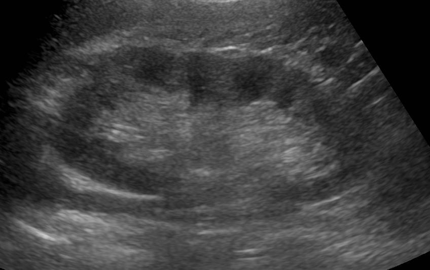
US – Increased Echogenicity
Ashley Davidoff TheCommonVein.net
46 year old female with discoid lupus erythematosus and renal failure. US shows increased echogenicity of the kidneys consistent with medical renal disease
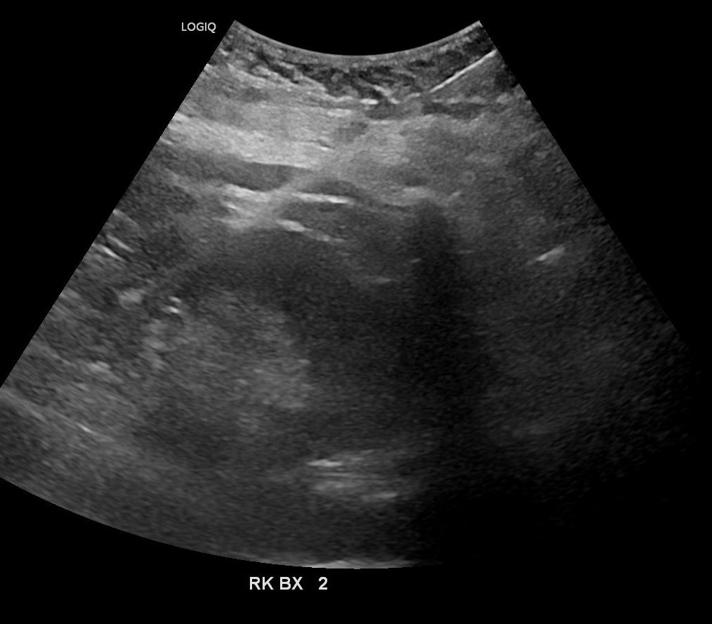
This study shows biopsy needle in the cortex of the right kidney
Ashley Davidoff TheCommonVein.net
Biopsy Result
Kidney biopsy which preliminarily is showing 60% sclerosis with evidence of chronic active interstitial nephritis
Post Biopsy Non Contrast CT
Ashley Davidoff TheCommonVein.net
Ashley Davidoff TheCommonVein.net
CT Shows Active Bleeding and Pseudoaneurysms
Post Biopsy Arterial Phase CT
Ashley Davidoff TheCommonVein.net
Ashley Davidoff TheCommonVein.net
Ashley Davidoff TheCommonVein.net
Ashley Davidoff TheCommonVein.net
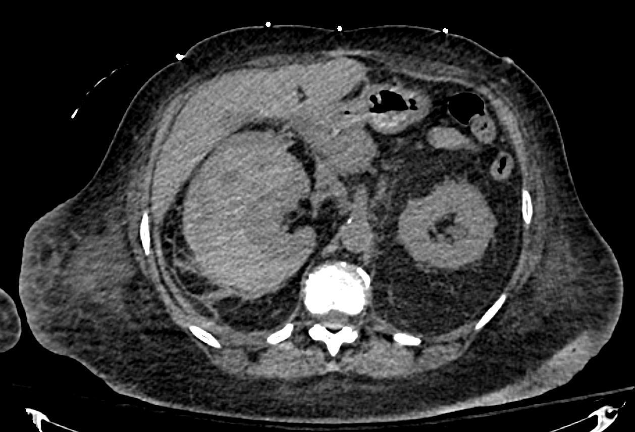
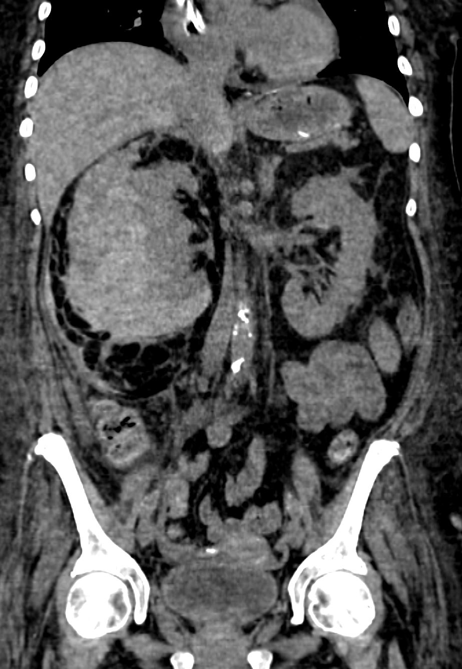
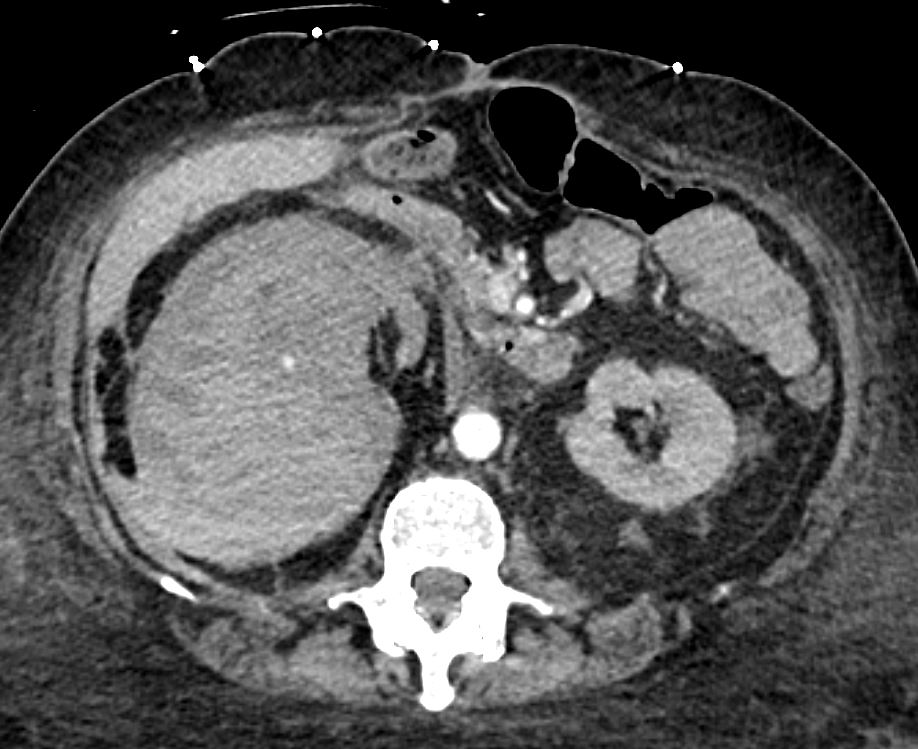
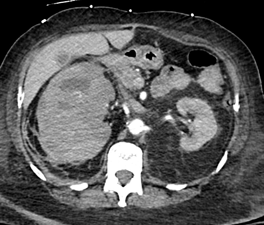
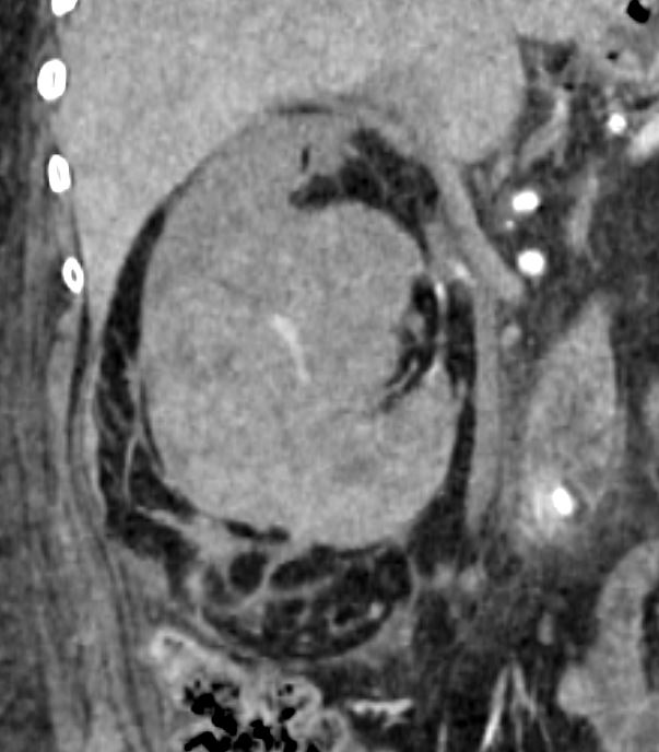
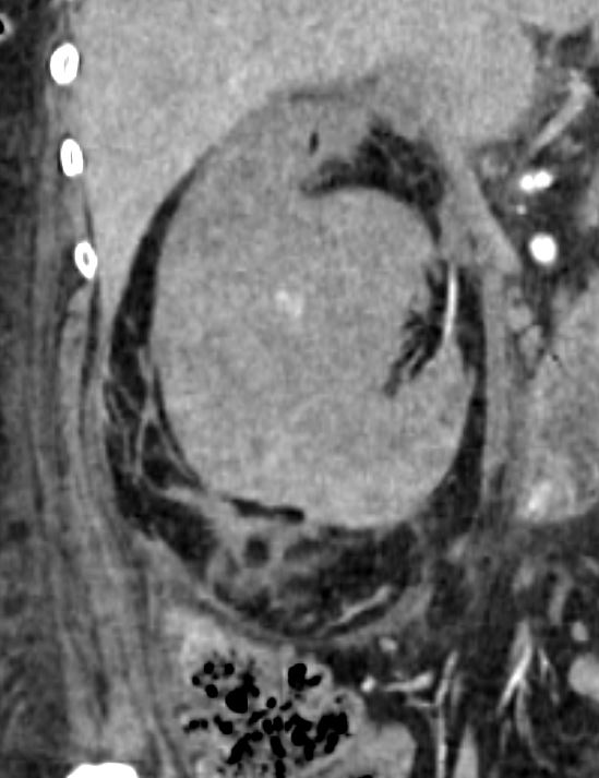
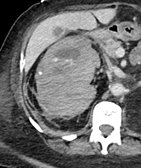
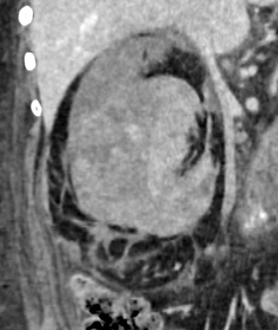
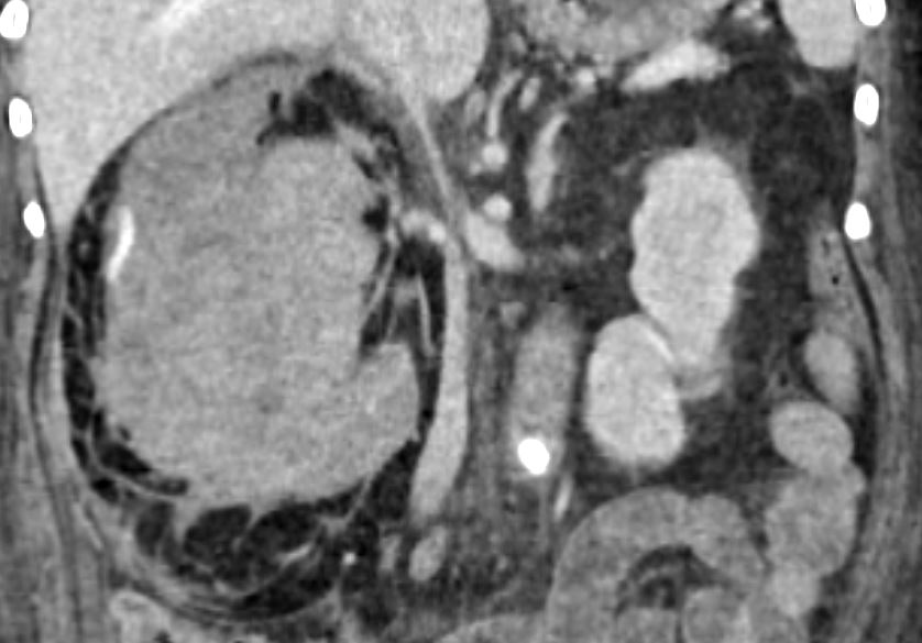
Post Biopsy Portal Venous Phase CT
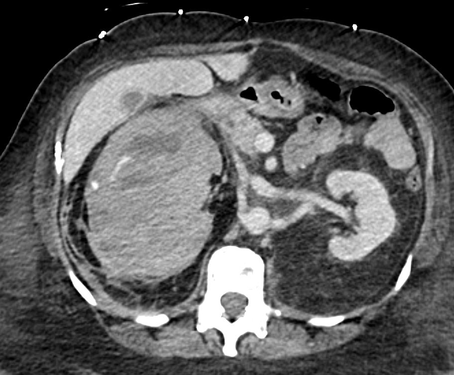
46 year old female with discoid lupus erythematosus and renal failure. CTscan during the portal venous phase post biopsy shows findings suggestive of pseudo-aneurysms and active bleeding with a large perinephric hematoma compressing the kidney
46 year old female with discoid lupus erythematosus and renal failure. CTscan during the portal venous phase post biopsy shows findings suggestive of pseudo-aneurysms and active bleeding with a large perinephric hematoma compressing the kidney
46 year old female with discoid lupus erythematosus and renal failure. CTscan during the portal venous phase post biopsy shows findings suggestive of pseudo-aneurysms and active bleeding with a large perinephric hematoma compressing the kidney
46 year old female with discoid lupus erythematosus and renal failure. CTscan during the portal venous phase post biopsy shows findings suggestive of pseudo-aneurysms and active bleeding with a large perinephric hematoma compressing the kidney
IR Angiography Multiple PSA’s and Active Bleeding
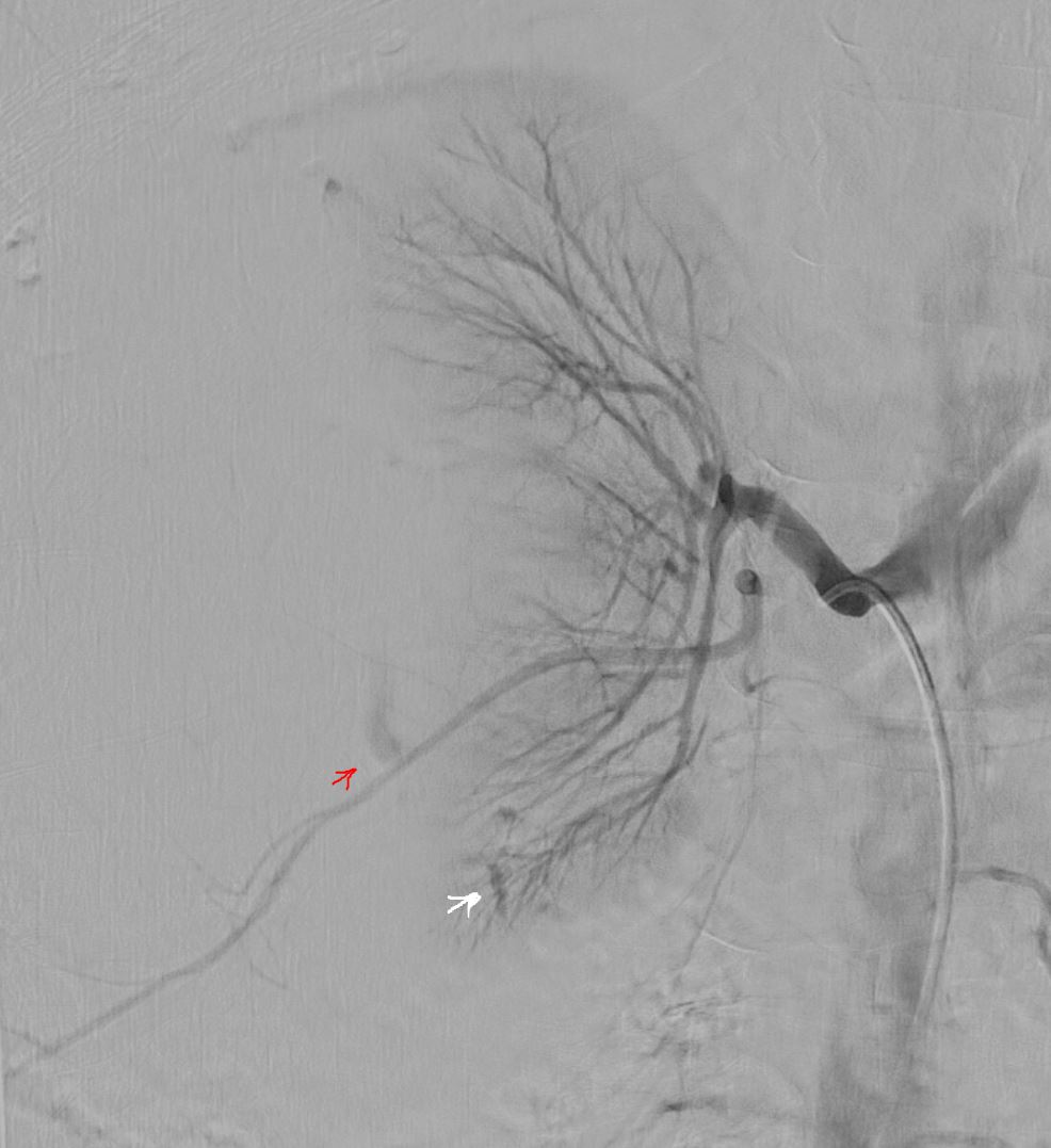
46 year old female with discoid lupus erythematosus and renal failure. Angiography injecting into the right renal artery post biopsy shows findings suggestive of pseudo-aneurysms (white arrow) and active bleeding (red arrow) with a large perinephric hematoma compressing the kidney
Ashley Davidoff TheCommonVein.net
Sub-selective IR Angiography Pseudoaneurysm
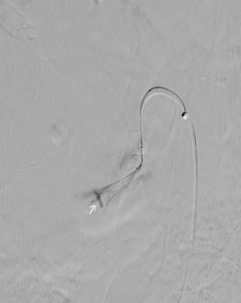
46 year old female with discoid lupus erythematosus and renal failure. Selective angiography injecting into the right renal artery post biopsy shows findings suggestive of pseudo-aneurysms and in a segmental artery in the lower pole
Ashley Davidoff TheCommonVein.net
IR Angiography Embolizations
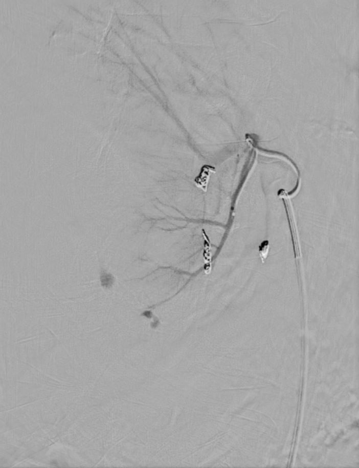
46 year old female with discoid lupus erythematosus and renal failure. Selective angiography injecting into the right renal artery post biopsy shows findings suggestive of pseudo-aneurysms and in a segmental artery in the lower pole Sub-selective catheterization with embolisation of 3 pseudoaneurysms and one active bleed
Ashley Davidoff TheCommonVein.net
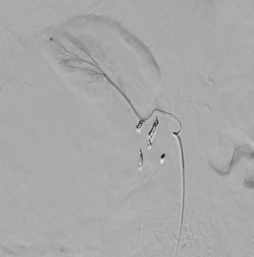
Final Result
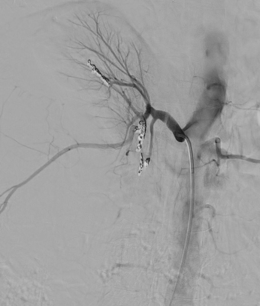
46 year old female with discoid lupus erythematosus and renal failure. Selective angiography injecting into the right renal artery post biopsy shows findings suggestive of pseudo-aneurysms and in a segmental artery in the lower pole Sub-selective catheterization with embolisation of 3 pseudoaneurysms and one active bleed
Ashley Davidoff TheCommonVein.net
Renal angiography demonstrates pseudoaneurysms involving three
subsegmental branches of the right renal artery and active contrast
extravasation from a fourth subsegmental branch. Selective catheterization
and coil embolization performed of each subsegmental branch to stasis.
Post embolization right renal angiography showed resolution of the
pseudoaneurysms and areas of active contrast extravasation. Mass effect
upon the right kidney seen from known perinephric hematoma.