
Cysts on Ultrasound
Cysts on CT
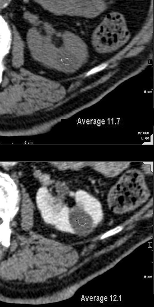
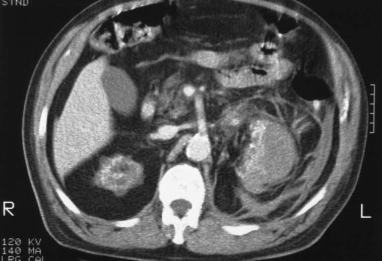
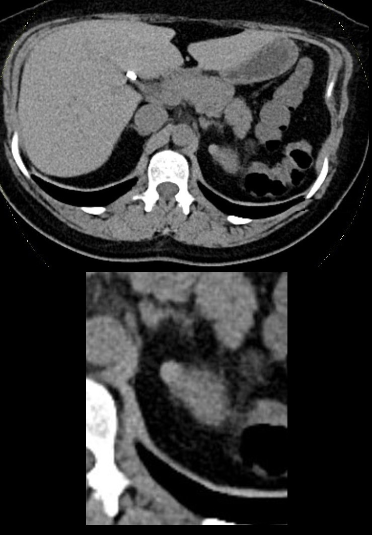
CT through the left kidney shows a low-density cyst prior to contrast measuring 11.7HU (upper image). Following contrast (lower image), the density of the nodule remained at 12 HU. The low density and lack of enhancement indicates a benign renal cyst
Ashley Davidoff MD TheCommonVein.net 135676
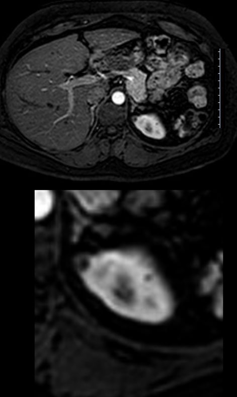
Cysts on MRI
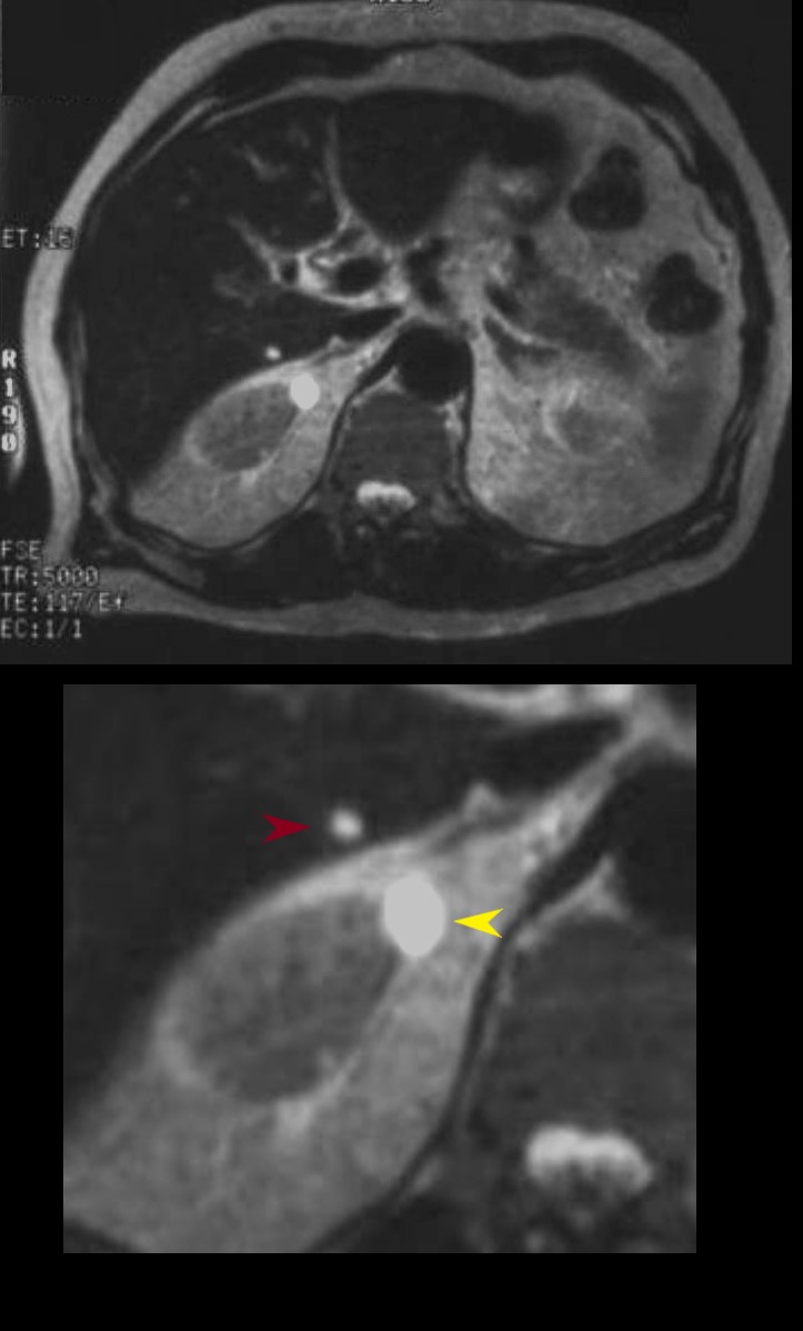
A T2 weighted MRI, shows a tiny well defined hyperintense unilocular cyst in the liver (maroon arrowhead) and a larger well defined hyperintense unilocular cyst exophytic cyst in the upper pole of the right kidney (yellow arrowhead) characteristic of benign disease
Ashley Davidoff MD TheCommonVein.net 02665
Cysts on CT
Congenital
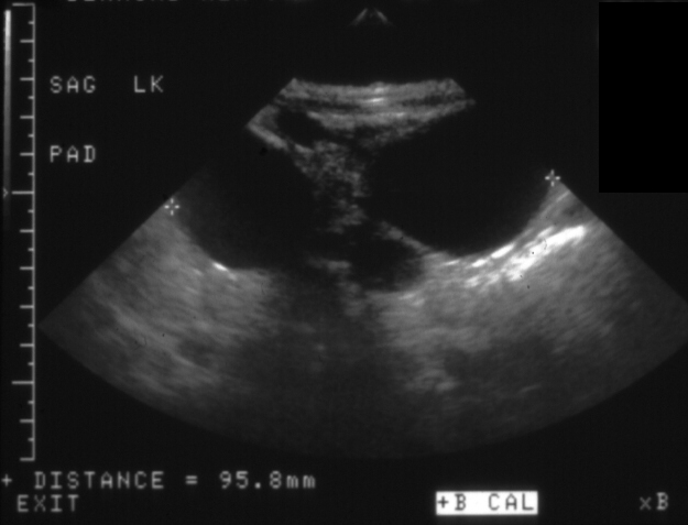
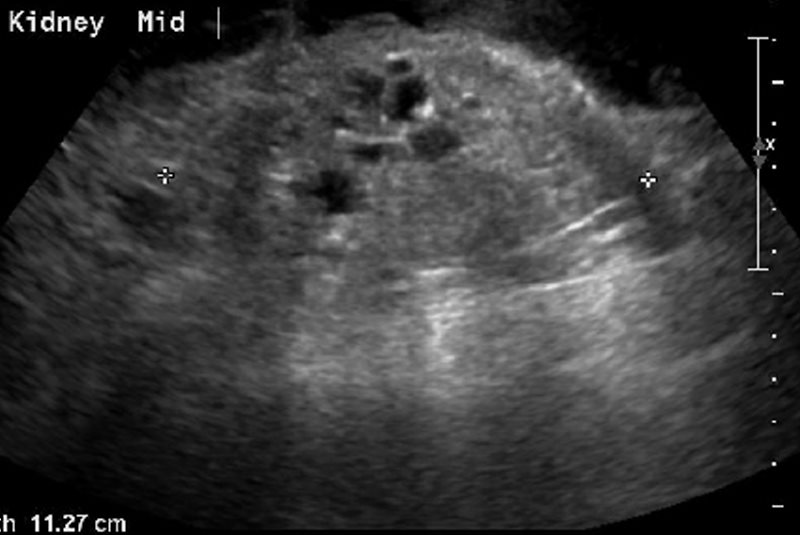
2 Month old presents with a abdominal mass and a cystic abnormality of the left kidney on ultrasound. US in the longitudinal plane shows multiple anechoic cysts of varying size, with evidence of backwall enhancement and through transmission. The kidney measures 9.6cms (normal at this age 4-5cms)
Final diagnosis was multicystic dysplastic left kidney and normal right kidney
Ashley Davidoff MD TheCommonVein.net See Case List 005K
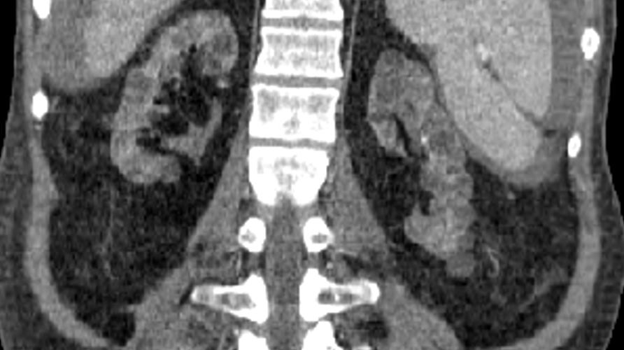
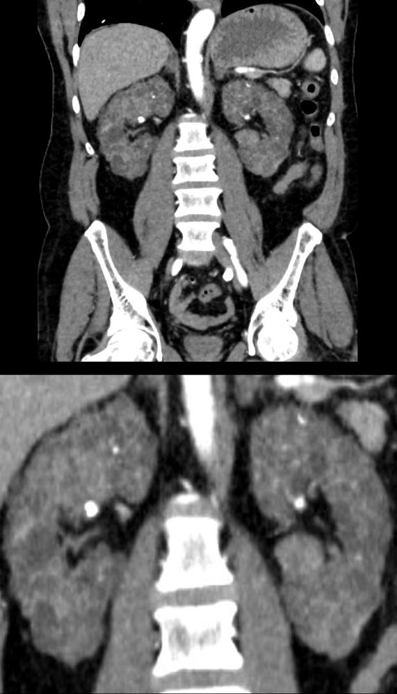
67 year old male with chronic renal failure. Reconstruction of a CT in the coronal plane shows bilateral diffuse small cysts consistent with the diagnosis of acquired cystic disease of chronic renal failure. Although the craniocaudal span is only mildly reduces there is significant parenchymal thinning and renal sinus lipomatosis. Noted ascites from peritoneal dialysis
Ashley Davidoff MD TheCommonVein.net
67 year old male with chronic renal failure. US in the longitudinal plane shows an 11.3cms echogenic kidney with multiple cysts in the 8mm to 10mms range consistent with the diagnosis of acquired cystic disease of chronic renal failure.
Ashley Davidoff MD TheCommonVein.net
Patient has acquired cystic disease of chronic failure and presents wit spontaneous subcapsular hemorrhage of the left kidney. A hemorrhage from a malignancy known to occur as a complication of this entity was suspected. At the inferior aspect of the hemorrhage there is extension of the process into the retroperitoneum giving the appearance of the “spider web sign” or “cobweb sign”
Ashley Davidoff MD TheCommonVein.net RnD
Polycystic Kidney Disease PCKD
56-year-old male in chronic renal failure presents with flank pain. CT with contrast shows diffuse cystic changes of near normal sized kidneys with total replacement of the renal parenchyma by small uniformly sized cysts. Multiple calcifications are scattered throughout the parenchyma.
Ashley Davidoff MD TheCommonVein.net 135723c
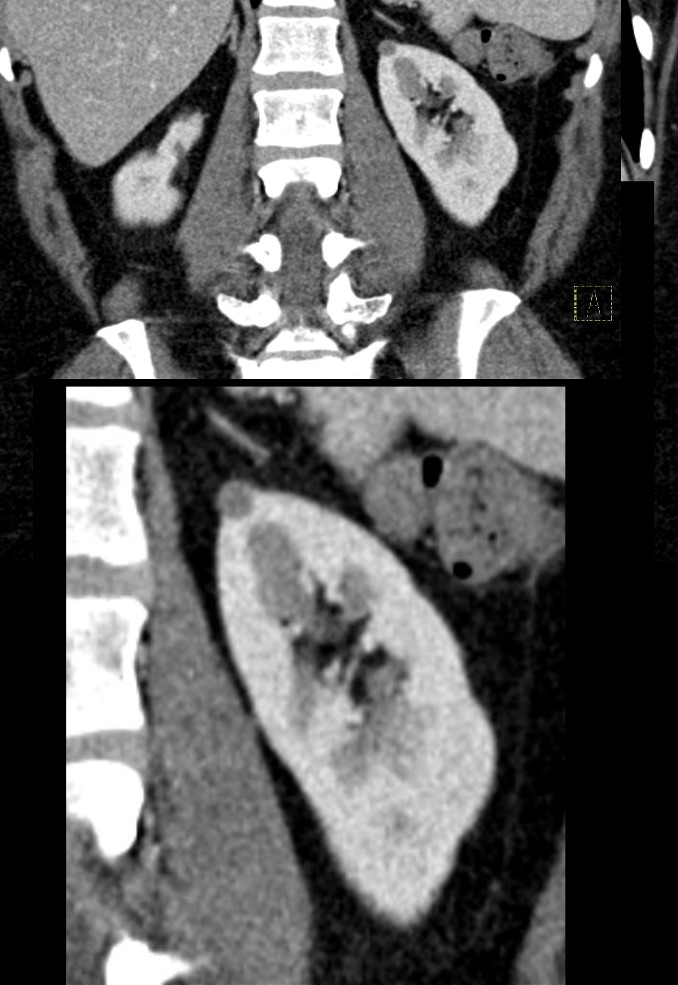
Hemorrhagic or Proteinaceous Cysts CT
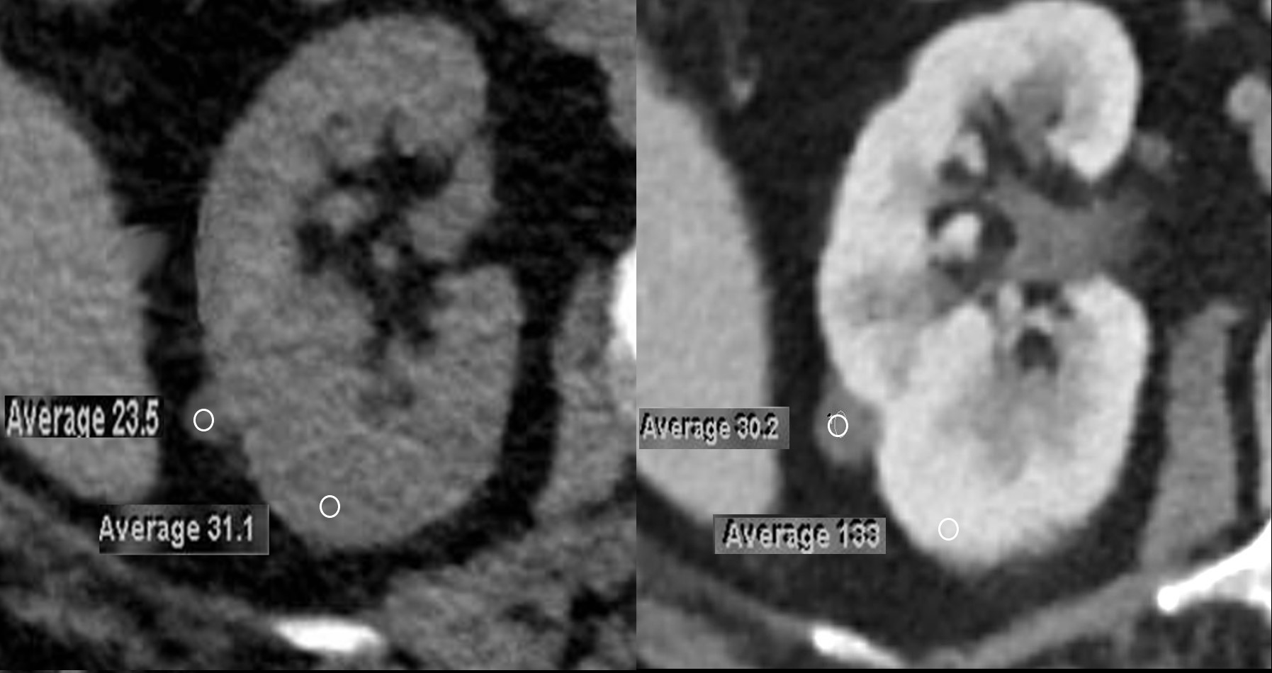
72-year-old female presents with abdominal pain. A 9mm homogeneously hyperdense cystic appearing lesion is noted on the non-contrast CT and it measures 23.5HU and is consistent with an hemorrhagic cyst or proteinaceous cyst. The lesion measures30HU following contrast (SD 10HU) in the late corticomedullary phase. The normal kidney measures 31.1HU before contrast and 133HU after contrast
Ashley Davidoff MD TheCommonVein.net 136019cL
Non-Contrast CT in the axial plane shows an 8mm homogeneous hyperdense lesion, exophytic off the upper pole of the left kidney. A hyperdense cyst as a result of hemorrhage or accumulation of proteinaceous material are most likely
Ashley Davidoff MD TheCommonVein.net TCV 24K 135914c
CT in the coronal plane shows an 8mm homogeneous that has now become relatively hypodense, exophytic off the upper pole of the left kidney. It does not appear to enhance.
A hemorrhagic or proteinaceous cyst is most likely. MRI subsequently confirmed the diagnosis
Ashley Davidoff MD TheCommonVein.net TCV 24K 135919c
Hemorrhagic or Proteinaceous Cysts MRI T1
T1 weighted MRI in the axial plane shows a T1 bright 8mm homogeneous lesion, exophytic off the upper pole of the left kidney.
This finding is compatible with a hemorrhagic or proteinaceous cyst
Ashley Davidoff MD TheCommonVein.net TCV 24K 135921c
Hemorrhagic or Proteinaceous Cysts MRI T2
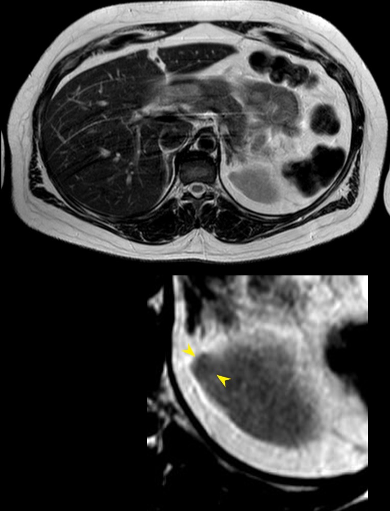
T2 weighted MRI in the axial plane shows a T1 dark 8mm homogeneous lesion, (yellow arrowheads lower image) exophytic off the upper pole of the left kidney confirming that the cyst contains protein or blood.
This finding is compatible with a hemorrhagic or proteinaceous cyst
Ashley Davidoff MD TheCommonVein.net TCV 24K 135925cL
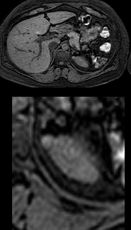
Hemorrhagic or Proteinaceous Cysts MRI Contrast
Contrast enhance MRI in the axial plane at 20seconds shows no enhancement of the matrix of the cyst. The wall does enhance but is thin.
This finding is compatible with a hemorrhagic or proteinaceous cyst
Ashley Davidoff MD TheCommonVein.net TCV 24K 135926c