
Neoplasm TCC
CT scan shows a 7mm filling defect on the anterior aspect of the right extrarenal pelvis . This was subsequently shown to be a transitional cell carcinoma
Ashley Davidoff MD TheCommonVein.net 135408
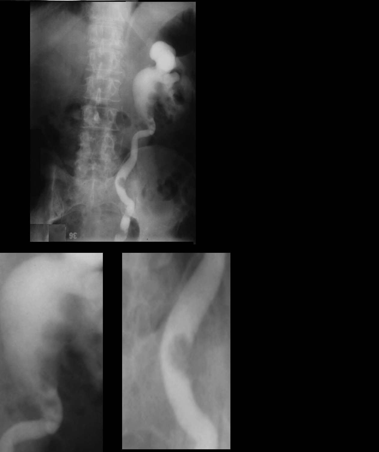
Retrograde study with injection of contrast into the left collecting system shows multilobulated filling defects in the dilated renal pelvis as well as filling defects in the proximal and mid ureter. The diagnosis was consistent with multicentric transitional cell carcinoma
Ashley Davidoff MD TheCommonVein.net
Key words kidney filling defect renal pelvis multicentric transitional cell carcinoma TCC
Ashley Davidoff MD TheCommonVein.net 06109
Filling Defect – Metabolic Stone
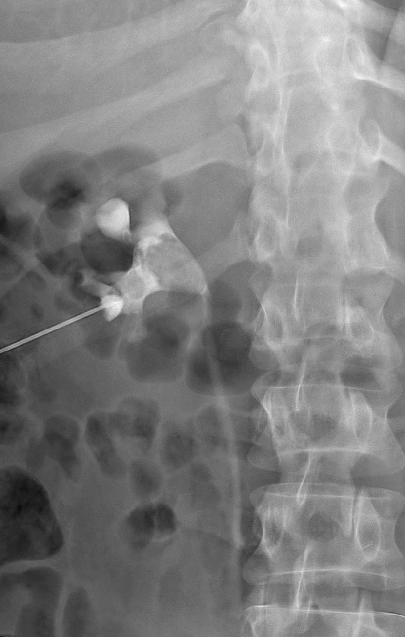
37year-old male presents with back pain Antegrade pyelogram shows 2 filling defects in the renal pelvis with the larger occupying almost the entire downstream pelvis. There is mild hydronephrosis. Contrast is seen in the ureter
Ashley Davidoff MD TheCommonVein.net 135460
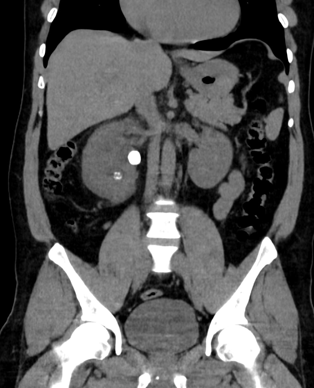
37year-old male presents with back pain Non contrast CT shows 2 calcifications. The larger stone is in the renal pelvis and the smaller in a lower pole calyx.
Ashley Davidoff MD TheCommonVein.net 135461