

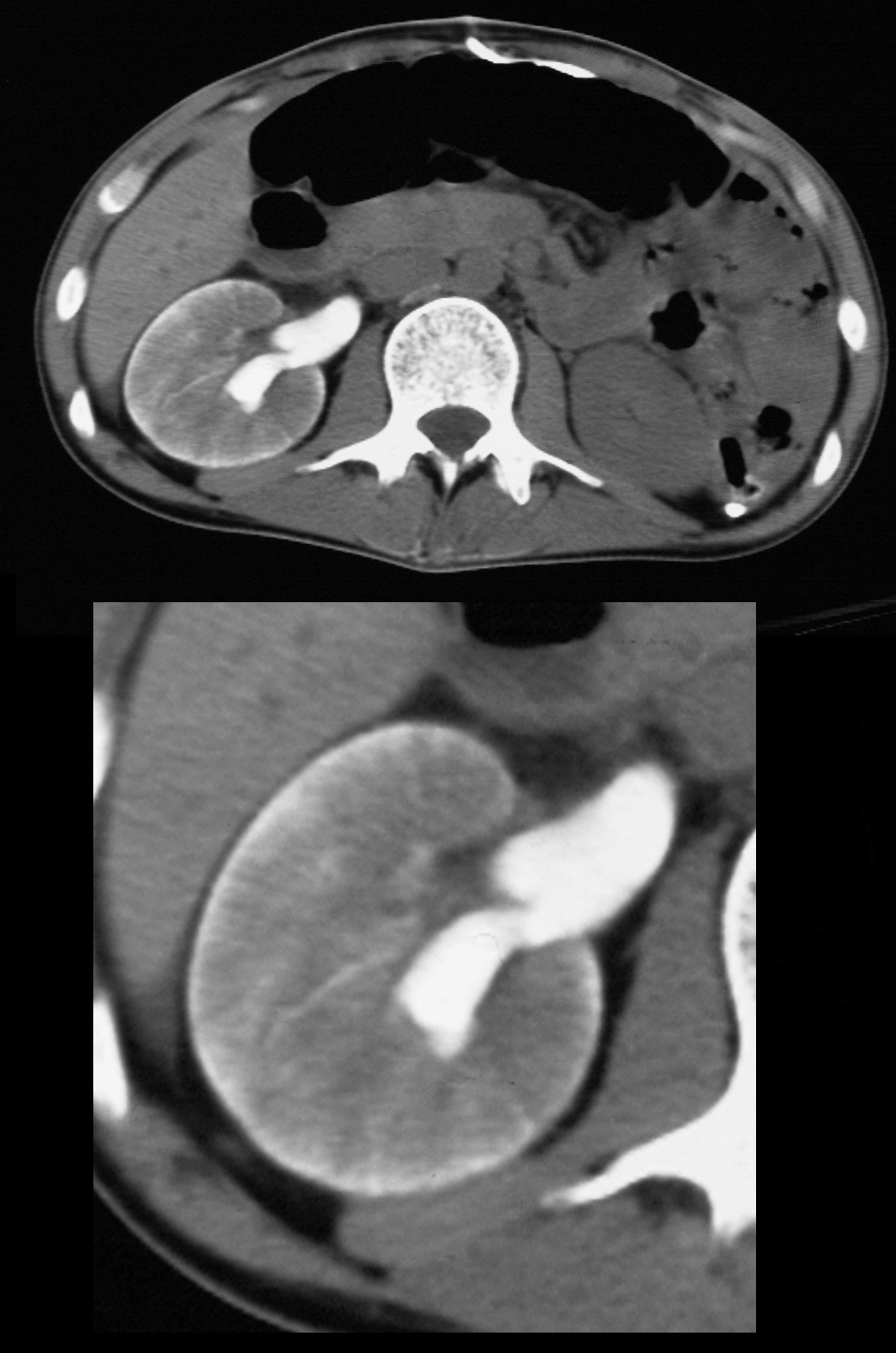
Axial CT scan in the pyelographic phase through the upper abdomen in a patient who presented with blunt trauma to the right kidney. There is a perfusion defect in the anterior aspect of the right kidney that involves both the cortex and medulla and extends to the capsular surface with evidence of thin rim of cortical enhancement, due to capsular perfusion. (“cortical rim sign”) and suggesting segmental arterial compromise.
Ashley Davidoff MD TheCommonVein.net 20267
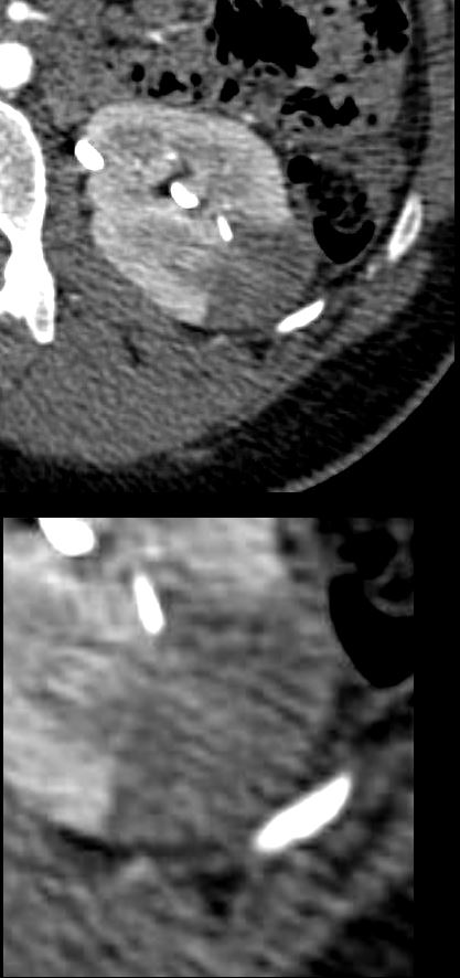
CT scan in the axial plane in a patient with flank pain shows a wedge-shaped swollen perfusion defect involving both the cortex and medulla of the left kidney and extending to the capsular surface. There is a hint of a capsular perfusion defect (cortical rim sign) .
Ashley Davidoff MD TheCommonVein.net
Ashley Davidoff MD TheCommonVein.net 40881