
44 year old male with Chronic Renal Failure s/p Renal Transplant and Osteodystrophy due to secondary hyperparathyroidism
keywords renal atrophy small kidneys renal sinus lipomatosis Ashley Davidoff TheCommonVein.net
CT scan shows renal transplant in the right lower quadrant
Ashley Davidoff TheCommonVein.net
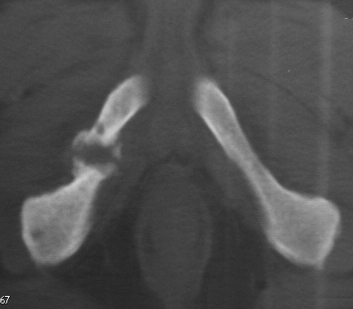
Scout film of a CT scan shows protrusio deformity in the right hip and a fracture
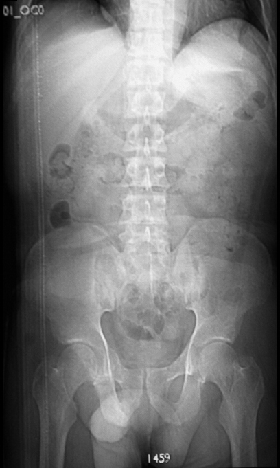
Scout film of a CT scan shows protrusio deformity in the right hip
Key words bone acetabulum dx osteomalacia kidney failure imaging radiology CTscan Ashley Davidoff TheCommonVein.net
Scout film of a CT scan shows protrusio deformity in the right hip
Key words bone acetabulum dx osteomalacia kidney failure imaging radiology CTscan Ashley Davidoff TheCommonVein.net
Brown tumors, also known as osteitis fibrosa cystica, are a rare manifestation of advanced primary or secondary hyperparathyroidism, which can be associated with renal osteodystrophy. They are not true tumors but rather localized, benign, bone lesions resulting from excessive parathyroid hormone (PTH) secretion and the subsequent resorption of bone. Brown tumors can affect various bones in the body, including those in the skull, long bones, ribs, and pelvis. When they occur in the context of renal osteodystrophy, it is often due to chronic kidney disease (CKD) and its associated abnormalities in mineral metabolism.
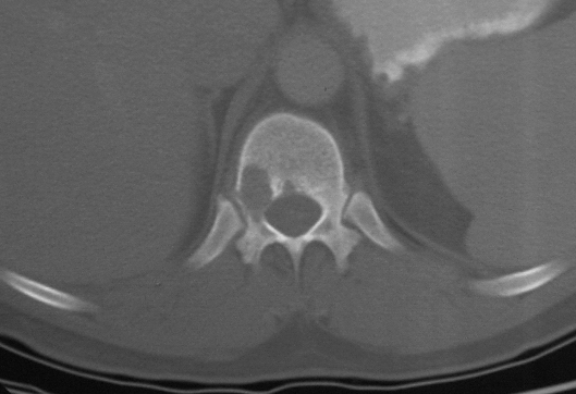
CT scan shows through the thoracolumbar spine shows a expansile lytic defect in the vertebral body with well-defined borders, consistent with a diagnosis of osteitis fibrosa lytica (brown tumor)
Key words bone acetabulum lytic defects osteitis fibrosa cystica brown tumors dx osteomalacia kidney failure imaging radiology CTscan Ashley Davidoff TheCommonVein.net
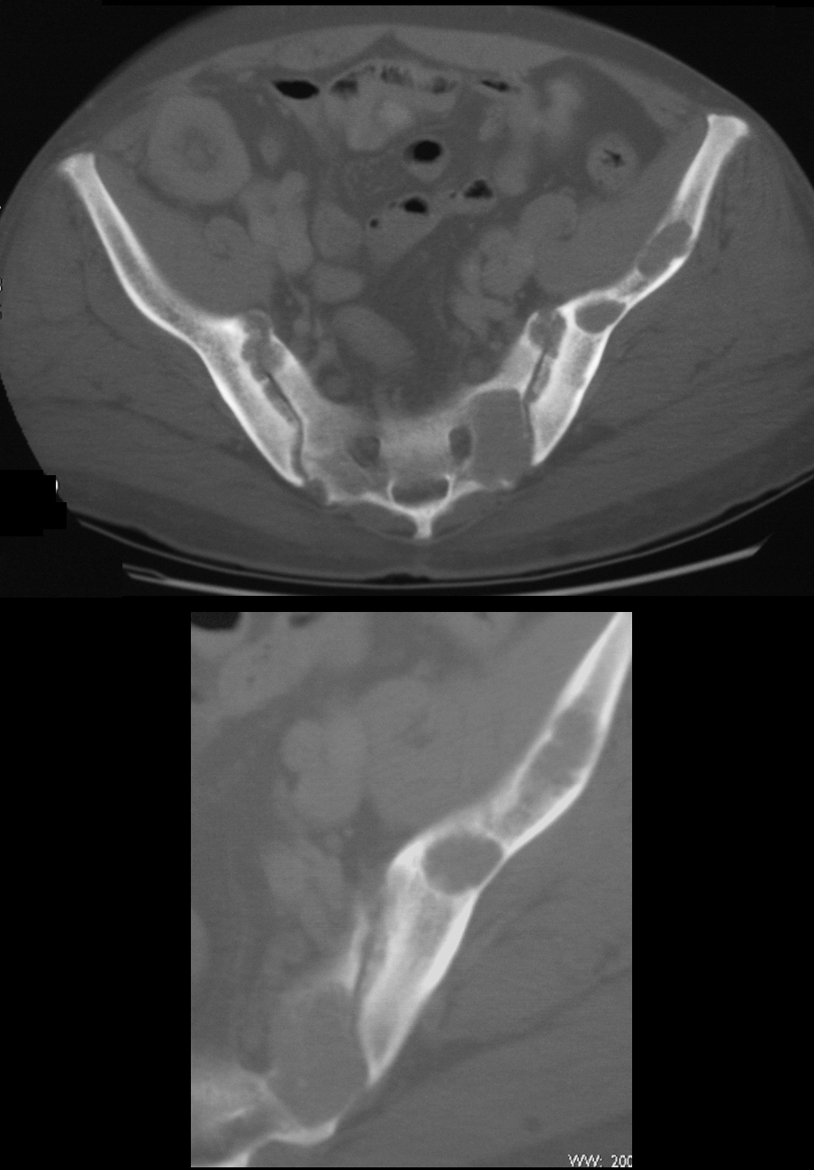
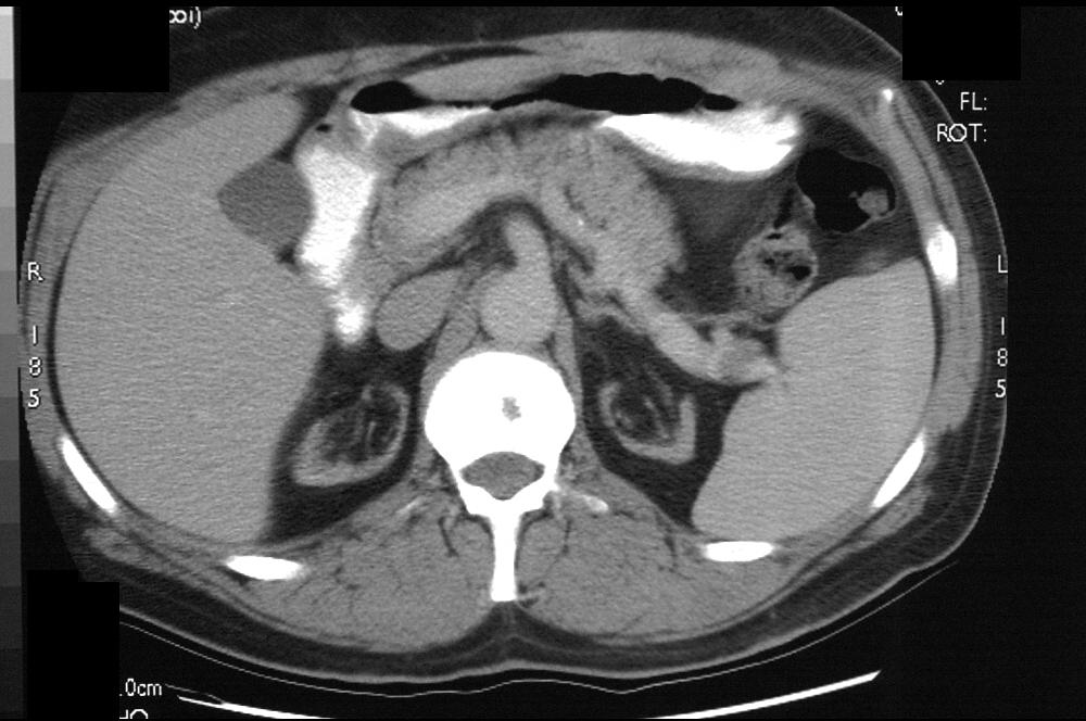
CT scan shows through the iliac bones shows multiple expansile lytic defects with well-defined borders in the iliac bones and sacrum, consistent with a diagnosis of osteitis fibrosa lytica (brown tumors)
Key words bone pelvis lytic defects osteitis fibrosa cystica brown tumors dx osteomalacia kidney failure secondary hyperparathyroidism imaging radiology CTscan Ashley Davidoff TheCommonVein.net
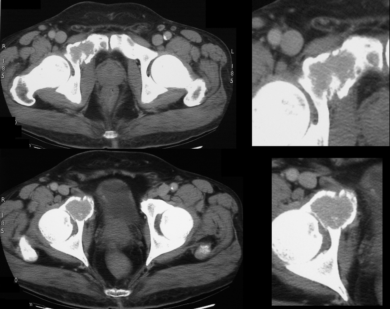
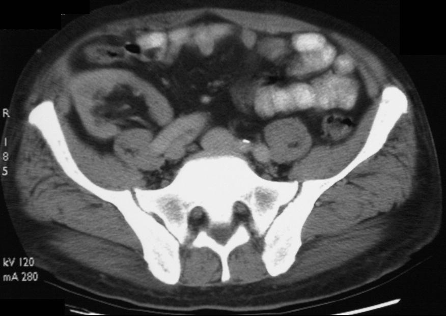
CT scan shows through the acetabula shows expansile lytic defect with well-defined borders in the right anterior column of the acetabulum extending into the superior pubic ramus consistent with a diagnosis of osteitis fibrosa lytica (brown tumor) There are multiple areas of cortical deficiency that could represent fractures
Key words bone acetabulum lytic defects osteitis fibrosa cystica brown tumors dx osteomalacia kidney failure secondary hyperparathyroidism imaging radiology CTscan Ashley Davidoff TheCommonVein.net
Looser’s Zones
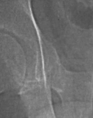
CT scan shows through the inferior pubic ramus shows transverse linear expansile region in the right inferior pubic ramus consistent with a diagnosis Looser’s zone
Key words bone pubic symphysis osteitis fibrosa cystica dx osteomalacia kidney failure secondary hyperparathyroidism imaging radiology CTscan Ashley Davidoff TheCommonVein.net
Looser’s zones, also known as Looser’s lines or pseudofractures, are radiographic findings associated with osteomalacia. Osteomalacia is a condition characterized by the softening and weakening of bones due to a deficiency in vitamin D, calcium, or phosphate. Looser’s zones represent areas of incomplete stress fractures or microfractures in bones and are typically seen on X-rays. These pseudofractures can be mistaken for actual bone fractures, but they are a result of weakened bone rather than acute trauma.