
Med Students
Cortical Phase
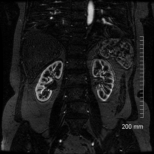
Cortical Phase
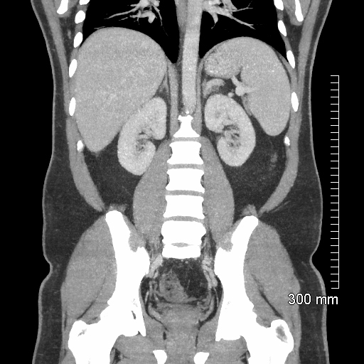
Corticomedullary Phase
Nephrographic Phase
Excretory Phase
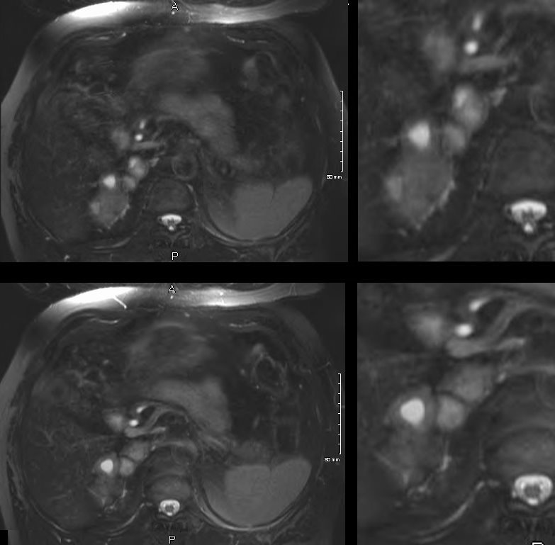
T1 Weighted Imaging
Pathology revealed a diagnosis of a benign Schwannoma
Ashley Davidoff MD TheCommonVein.net
T2 Weighted Imaging
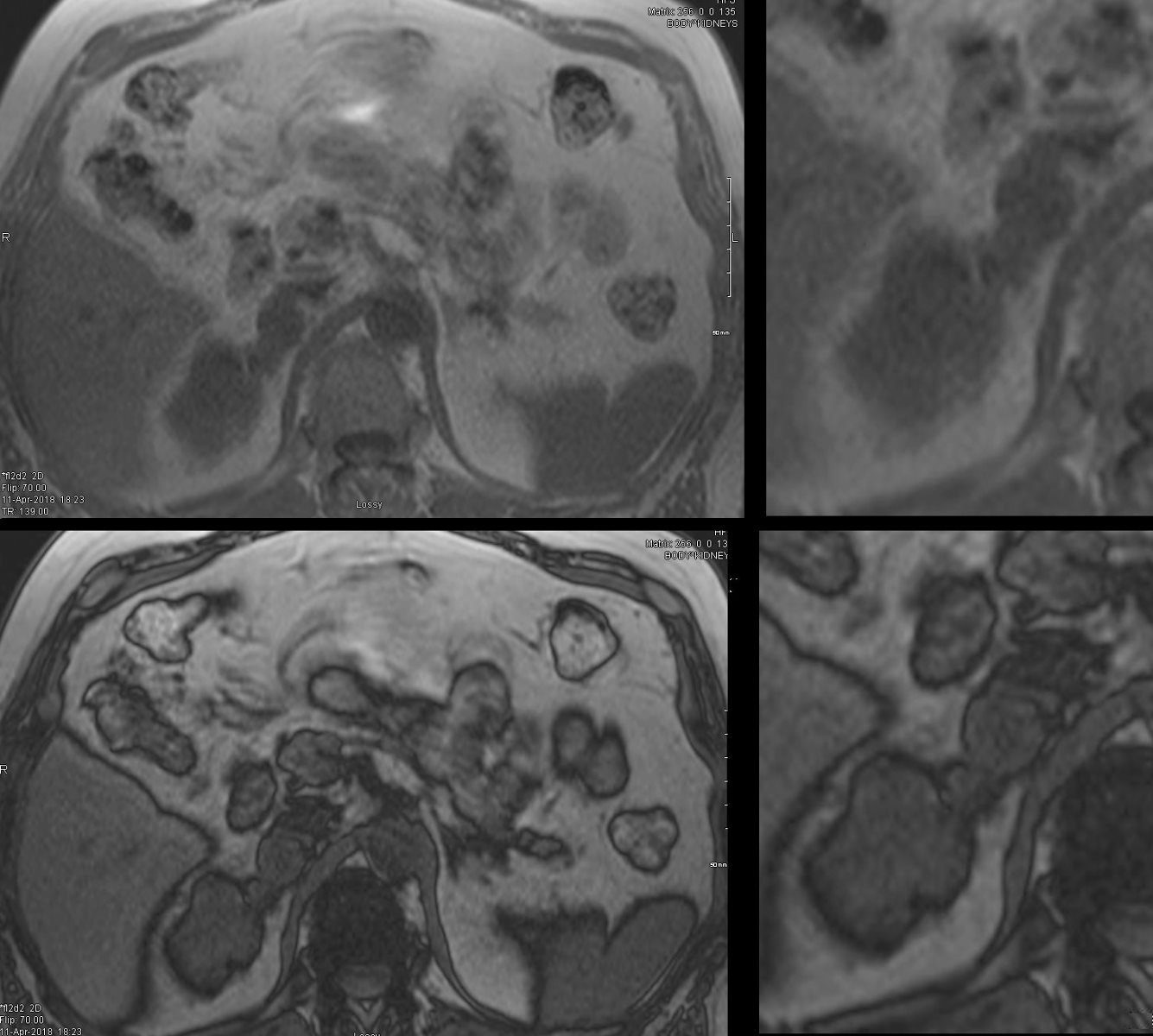
 T2 weighted MRI of a 58 year old male lobulated mass originating from the upper pole of the right kidney showing components of T2 brightening . The mass shows a combination of T2 brightness . In the nodule closest to the superior pole of the kidney the central portion (best seen in the right lower image) is as bright as the CSF and likely cystic in nature .It is associated with a thick wall. The nodular components abutting the vertebral are less bright and less well defined. Pathology revealed a diagnosis of a benign Schwannoma
Ashley Davidoff TheCommonVein.net

Ashley Davidoff TheCommonVein.net
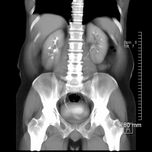
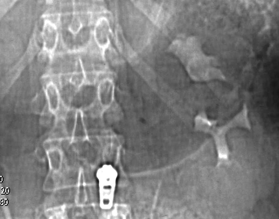
55 year old female presents with abdominal pain Scout film following contrast administration shows bilateral excretion with mass effect in the calyces of the midportion of the left kidney. with question of mild hydronephrosis (widening of the forniceal angle) of the left upper pole compound calyx. Ureters and bladder appear normal except for mild mass effect on the superior aspect of the right side of the bladder likely from the uterus
Diagnosis left sided renal cyst
Ashley Davidoff MD TheCommonVein.net RnD
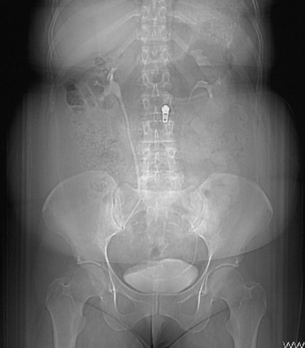
55 year old female presents with abdominal pain Focus on the upper pole of the left kidney on the scout film following contrast administration confirms the mass effect on the calyces in the midportion of the left kidney. And mild hydronephrosis (widening of the forniceal angle) of the left upper pole compound calyx. The lower pole calyces are normal
Diagnosis left sided renal cyst
Ashley Davidoff MD TheCommonVein.net RnD 006K
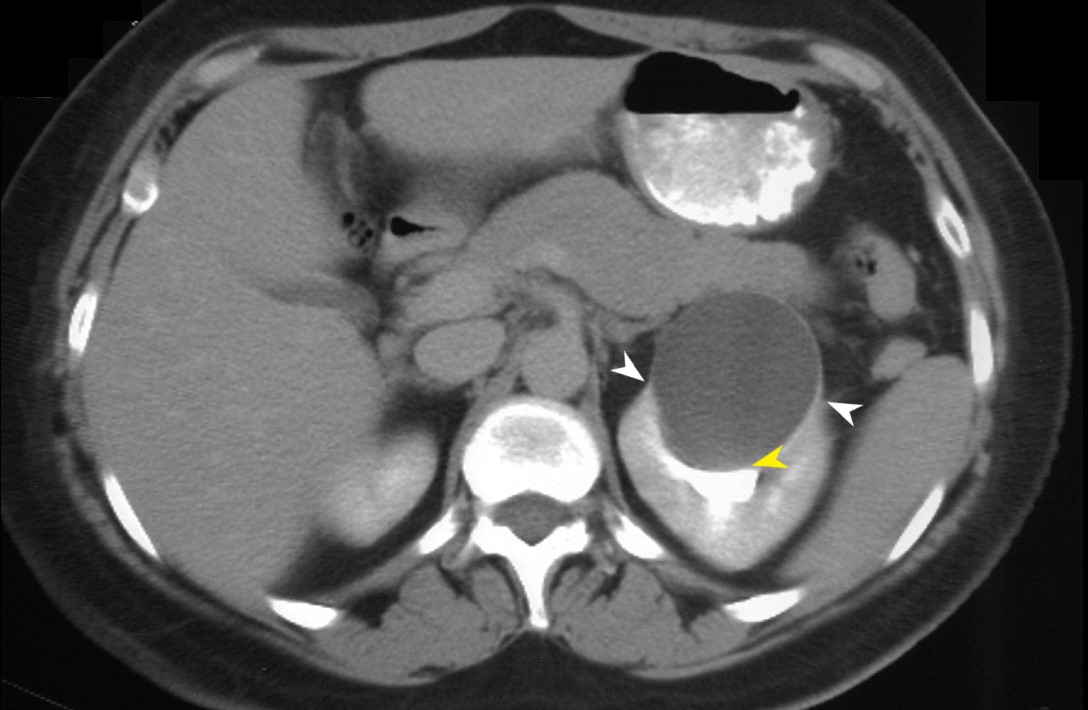
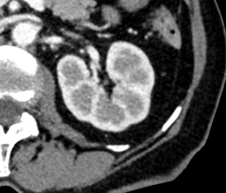
55-year-old female presents with abdominal pain. An axial CT scan through the upper portions of the left kidney shows distortion and compression of the upper pole calyces (yellow arrowhead) . There are bilateral “beak signs” (white arrowheads) indicating a benign slow growing cyst
Diagnosis: Left Sided Simple Renal Cyst Bosniak 1
Ashley Davidoff MD TheCommonVein.net RnD 006K
Hemorrhage
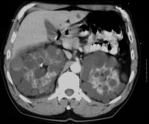
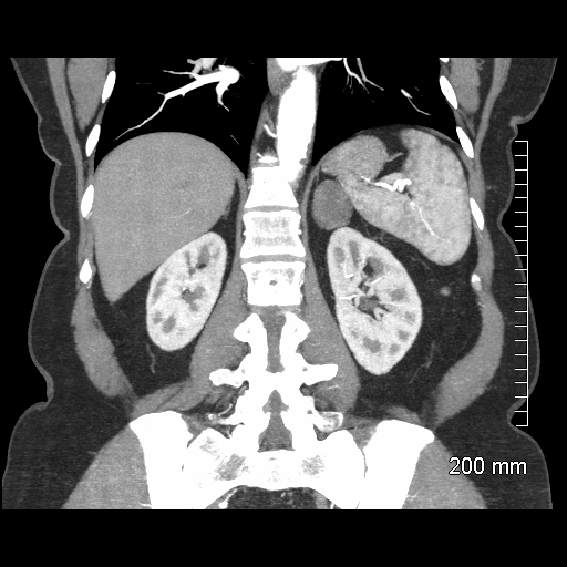
CT scan with contrast of a 54 year old male with polycystic kidney disease. In this case, the cysts are distributed in a relatively orderly and homogeneous fashion in the periphery of the both kidneys with an unusual symmetry. There is a reasonable amount of functioning parenchyma. One of the cysts in the periphery of the right kidney has a higher density and high density sediment suggesting hemorrhage
Ashley Davidoff MD TheCommonVein.net
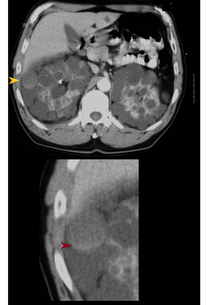
CT scan with contrast of a 54 year old male with polycystic kidney disease. In this case, the cysts are distributed in a relatively orderly and homogeneous fashion in the periphery of the both kidneys with an unusual symmetry. There is a reasonable amount of functioning parenchyma. One of the cysts in the periphery of the right kidney (yellow arrowhead) has a higher density and high density sediment (red arrowhead lower image suggesting hemorrhage
Ashley Davidoff MD TheCommonVein.net