
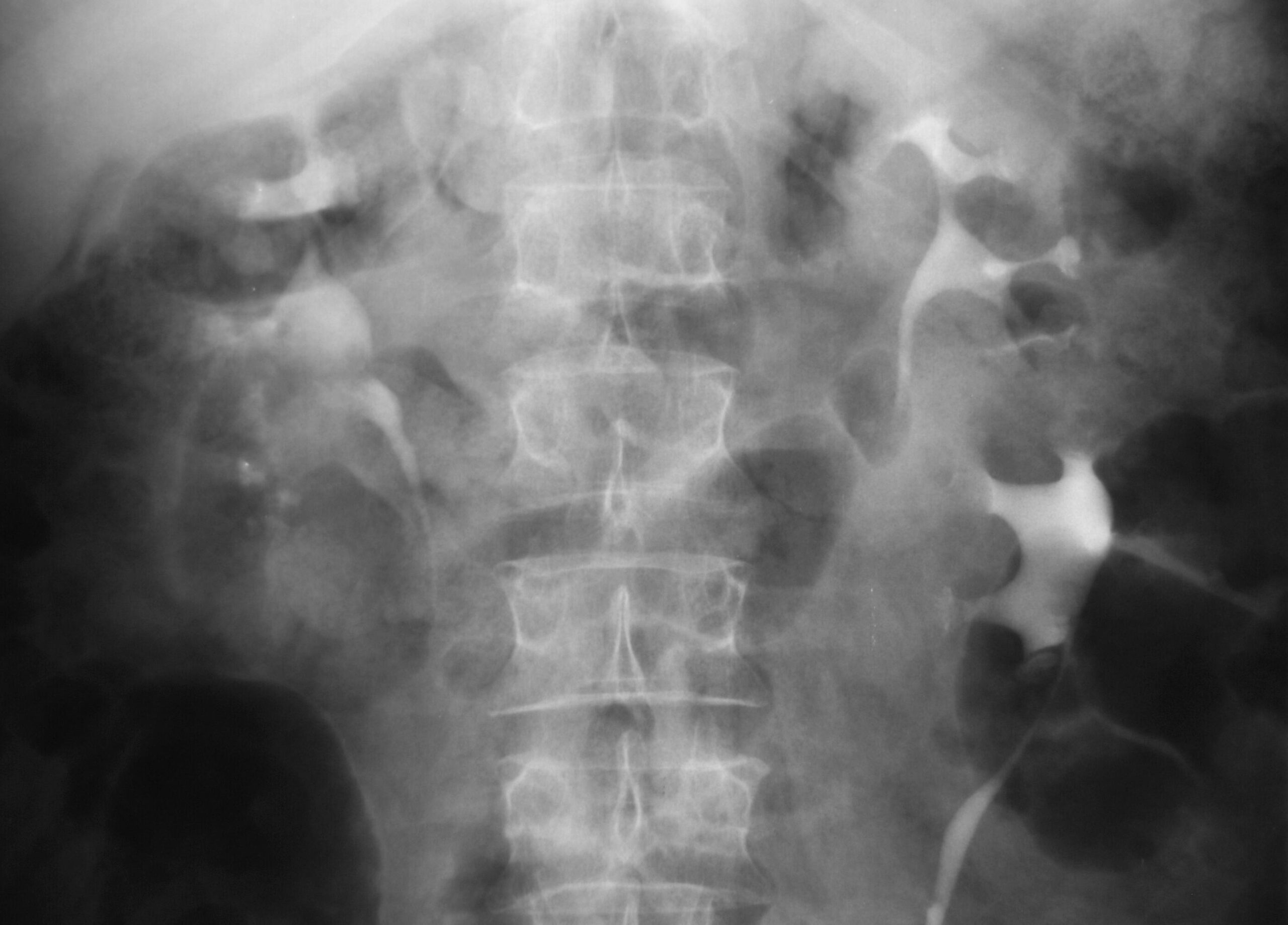
Duplication
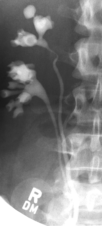
Excretory phase shows duplication of the upper and lower pole moieties of the left kidney. The two systems join at the renal pelvis and continue as one ureter to the bladder
Ashley Davidoff MD TheCommonVein.net 130887
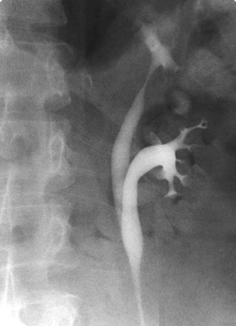
Excretory phase shows duplication of the upper and lower pole moieties of the right kidney. The two systems travel separately into the bladder. Incidental note is made of calyceal diverticulum arising from the upper pole moiety
Ashley Davidoff MD TheCommonVein.net 126115b
Bilateral Malrotation
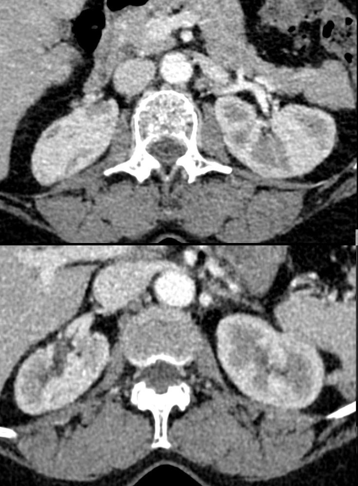
CT with contrast in the axial plane in a 63-year-old female shows malrotation of both kidneys The left kidney has its’ hilum pointing anteriorly (top image), and the right kidney has its’ hilum pointing laterally (bottom image)
Ashley Davidoff MD TheCommonVein.net 135727
Horseshoe Kidney
CT with contrast, reconstructed in the coronal plane a horseshoe kidney draped over the psoas muscles,.
Ashley Davidoff MD TheCommonVein.net 133211
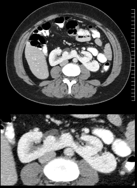
Pelvic Kidney
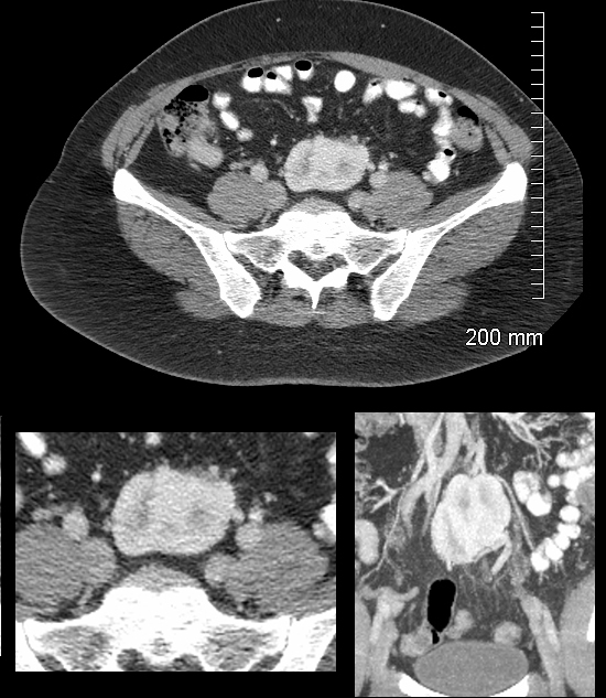
CT in the axial plane shows a reniform structure in the presacral region between the cortico-medullary and nephrographic phase of the contrast enhanced study. The bottom left image is a magnification view of the axial CT. The bottom right image is in the coronal plane and shows a mal-oriented pelvic kidney in the same recognizable phase of renal contrast excretion. A normal kidney was noted in the right renal fossa and the diagnosis is consistent with a pelvic kidney
Ashley Davidoff MD TheCommonVein.net 132055c
Crossed Fused Ectopia
Excretory phase shows cross fused ectopia on the left resulting in 2 fused kidneys, each with an independent ureter, and a separate independent normal appearing kidney on the right. This person has “3” kidneys
Ashley Davidoff MD TheCommonVein.net 40881
Ureteroceles
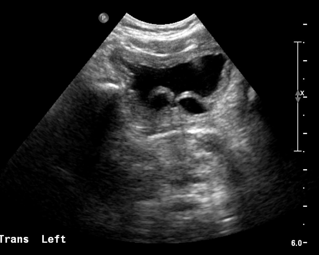
.Ultrasound of the bladder in the transverse projection in an 8-week-old infant male, shows bilateral cystic structures in the region of the UVJ (ureterovesical junctions) consistent with bilateral ureteroceles (cobra head x 2).
Ashley Davidoff MD TheCommonVein.net 135444