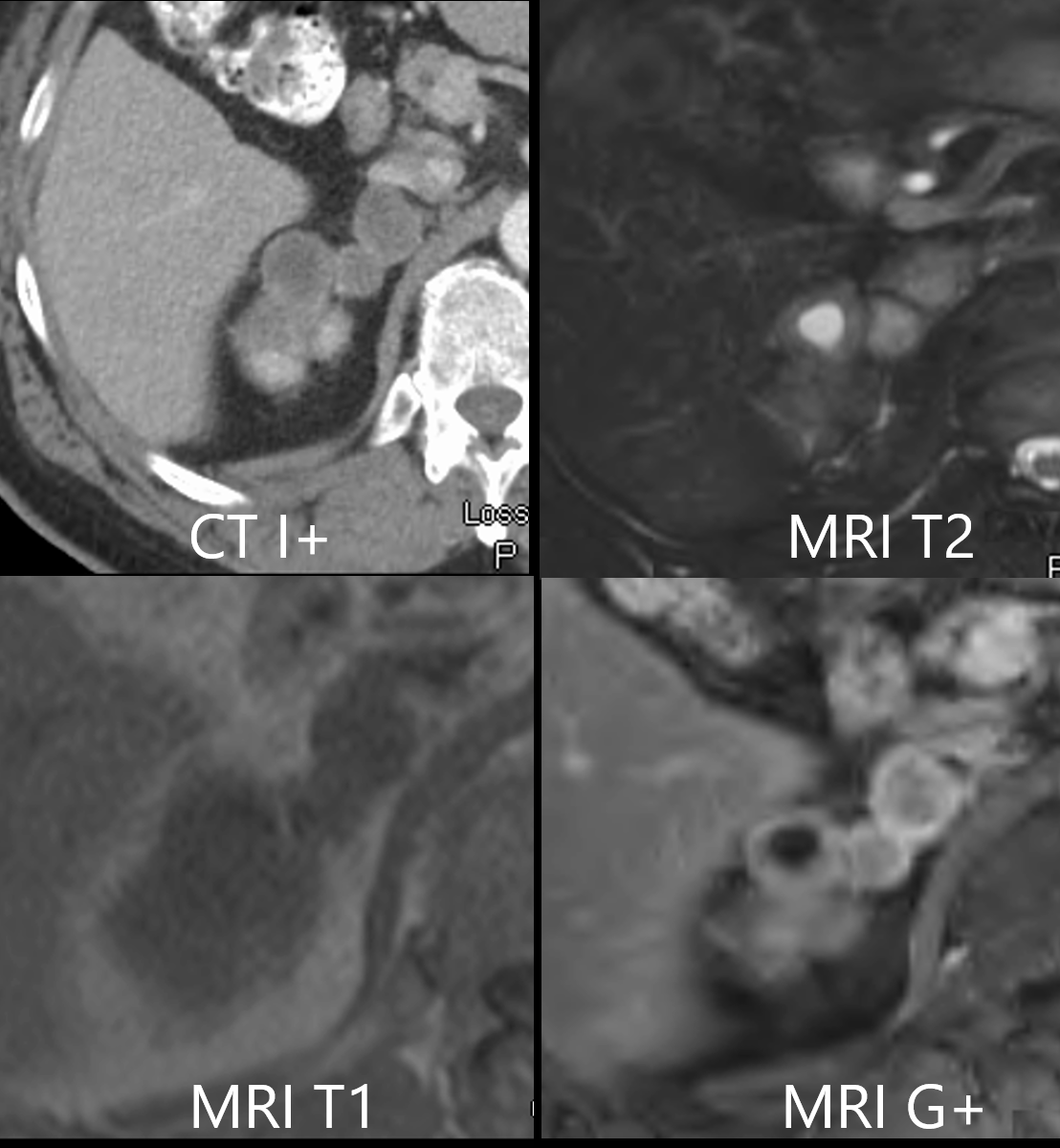
Collage of a axial imaging through a right upper pole mass in a 58 year old male showing a lobulated mass, varicoid in shape. using CT and MRI The left upper image shows a contrast enhanced CT with relatively low density matrix (41HU) and enhancing thick smooth wall. A T2 weighted image (left upper) shows a combination of T2 brightness and intermediate signal. The left lower image is a T1 weighted image showing isointensity of the mass with other soft tissues while the contrast enhanced MRI shows enhancing walls (41HU) lobulated mass originating from the upper pole of the right and a combination in the matrix of the lobules of dark signal and intermediate signal. Pathology revealed a diagnosis of a benign Schwannoma Ashley Davidoff MD TheCommonVein.net