
Sickle Cell Disease – Anemia
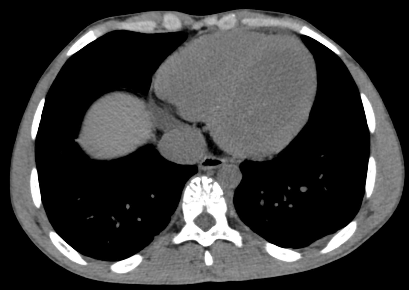
CT without contrast in the axial plane in a 23-year-old male shows low density blood in the left ventricular cavity enabling the visualization of the LV myocardium. This is due to the anemia of the patient which causes a decrease in the density of blood
Ashley Davidoff MD TheCommonVein.net 135731
Sickle Cell Disease Bilateral Microinfarcts and
Enlarged Left Kidney
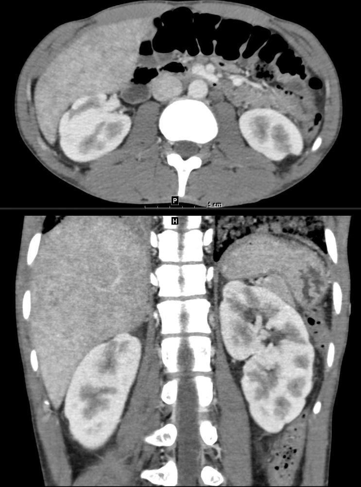
CT with contrast in the coronal plane in a 23-year-old male shows an enlarged left kidney measuring close to 13cms. with evidence of bilateral scarring attributed to microinfarcts from sickle cell disease. Note the stomach has taken up the space since the spleen was almost totally infarcted.
Ashley Davidoff MD TheCommonVein.net 135728
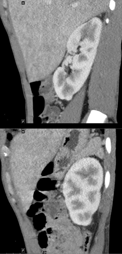
CT with contrast in the sagittal plane plane in a 23-year-old male shows an enlarged left kidney measuring close to 13cms in the upper panel and the normal sized kidney in the lower panel. There is evidence of bilateral scarring attributed to microinfarcts from sickle cell disease
Ashley Davidoff MD TheCommonVein.net 135730
Sickle Cell Disease – Unusual Vascular Markings on the Vertebra
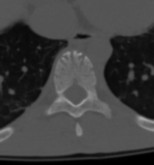
CT without contrast in the axial plane in a 23-year-old male shows unusually prominent markings on the anterior surface of the vertebral body possibly reflecting enlarged blood vessels
Ashley Davidoff MD TheCommonVein.net 135731
Links and References
Gardiner K The Renal Sonographic Appearance of Sickle Cell Disease JDMS 3:14 -19 Jan 1987