
Pathology
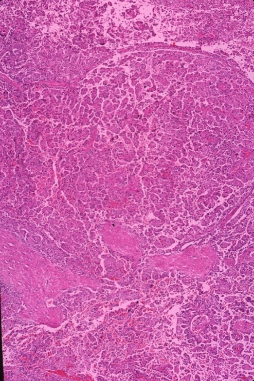
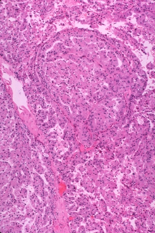
Histology of Renal Cell Carcinoma
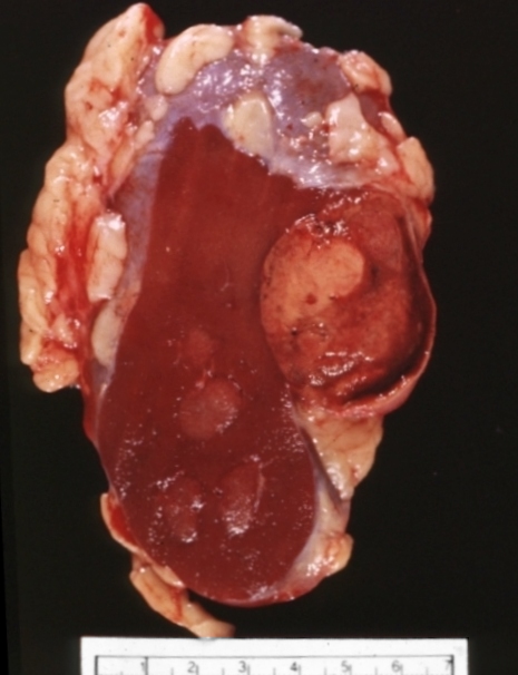
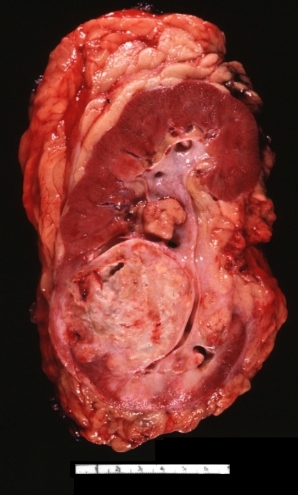
Clear Cell Type
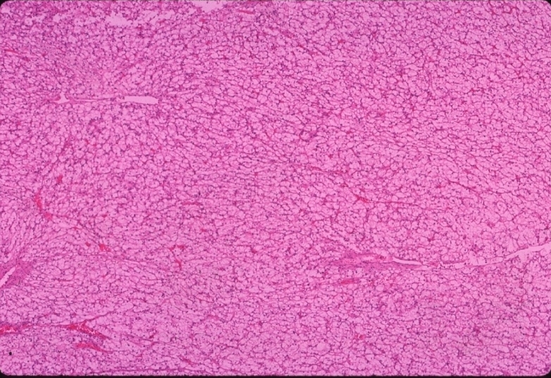
Ashley Davidoff MD
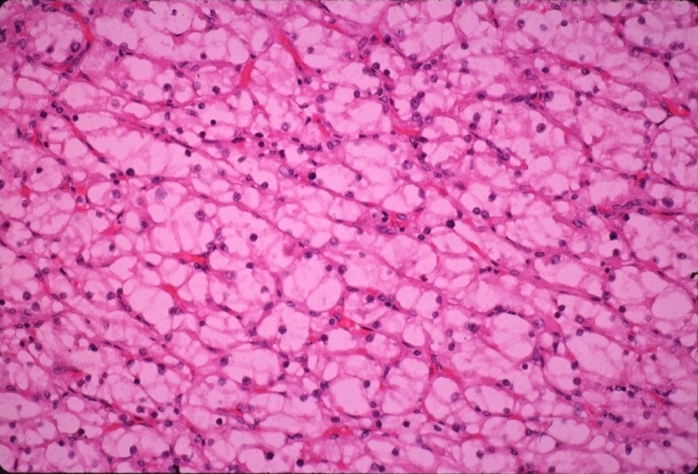
Ashley Davidoff MD
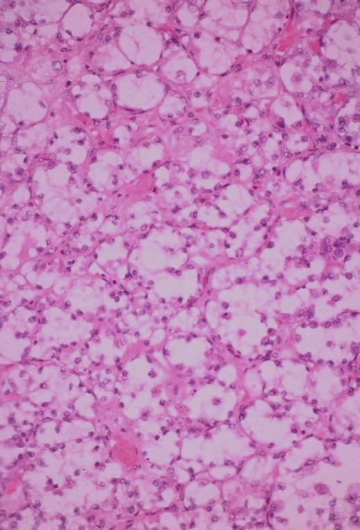
Ashley Davidoff MD
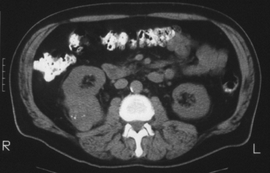
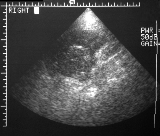
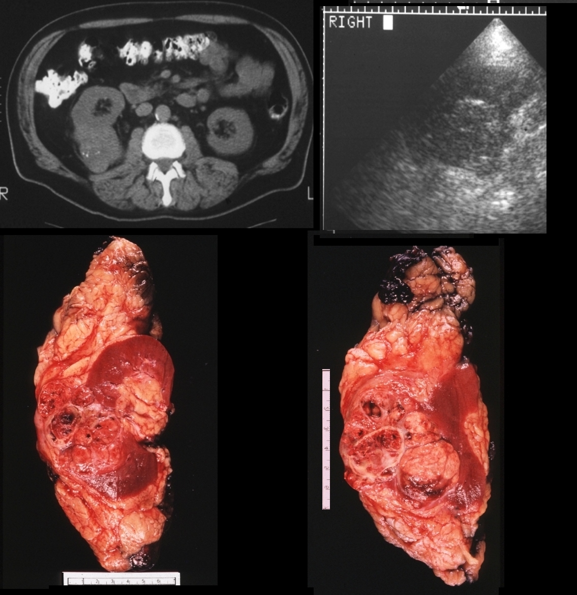
Papillary Cell Type RCC
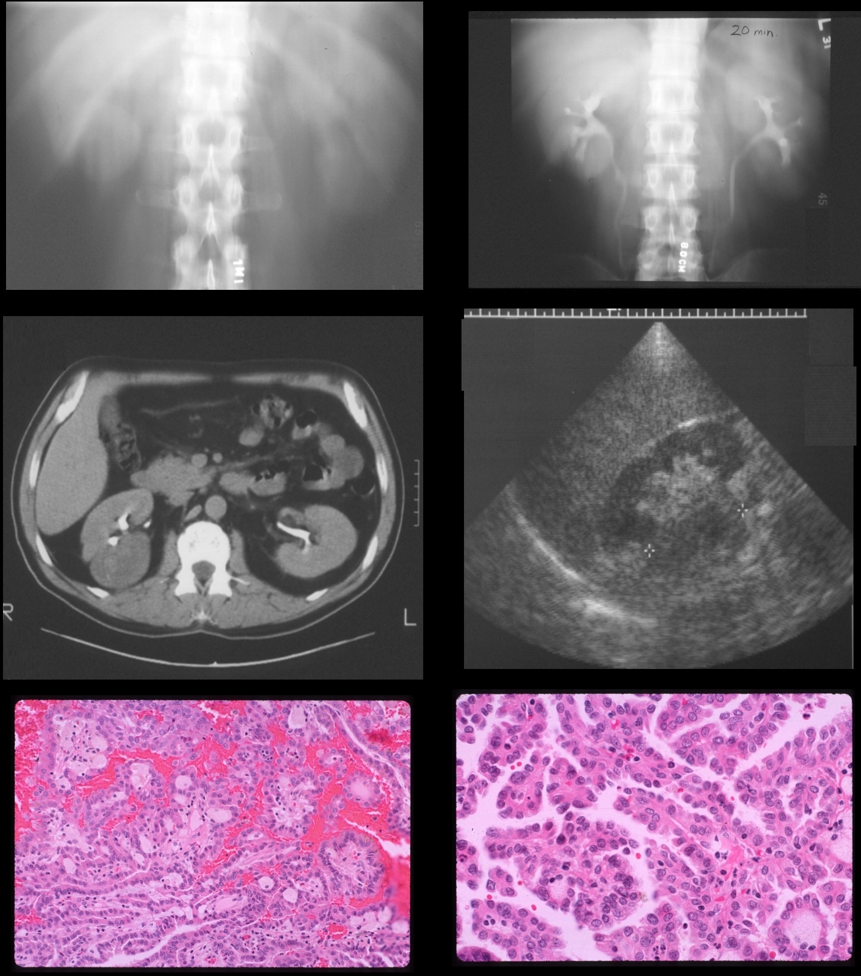
64 year old male
IVP at 20 seconds shows a mass in the right lower pole and 20 minute film conforms the finding.CT shows a deforming mass and US shows mildly hypoechoic mass. Pathology reveals papillary cell RCC
Ashley Davidoff MD
Granular cell Type
Ashley Davidoff MD

Ashley Davidoff MD
Renal Cell Carcinoma with Suggestion of Extension Beyond the Renal Capsule
Ashley Davidoff MD
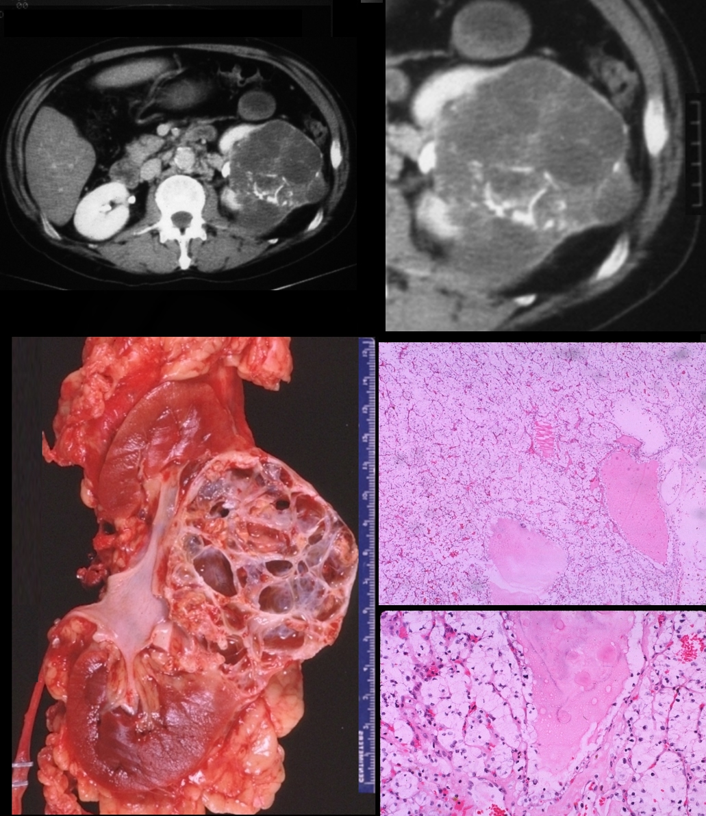
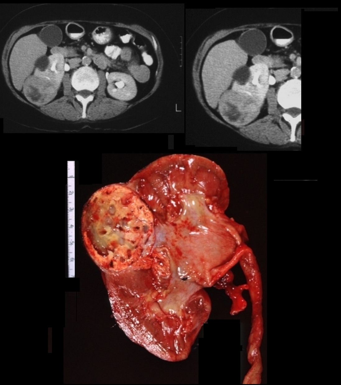
Cystic renal cell carcinoma with dystrophic calcification in the mid portion of the left kidney. Pathology reveals cystic renal cell carcinoma with clear cell histopathology
Ashley Davidoff MD
Ashley Davidoff MD
Ashley Davidoff MD
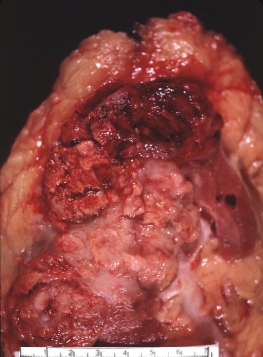
Extension beyond the renal capsule
Ashley Davidoff MD
Ashley Davidoff MD
On IVP
showing a mass in the right lower pole extending into the renal pelvis
Ashley Davidoff MD
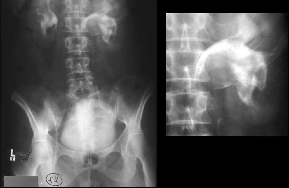
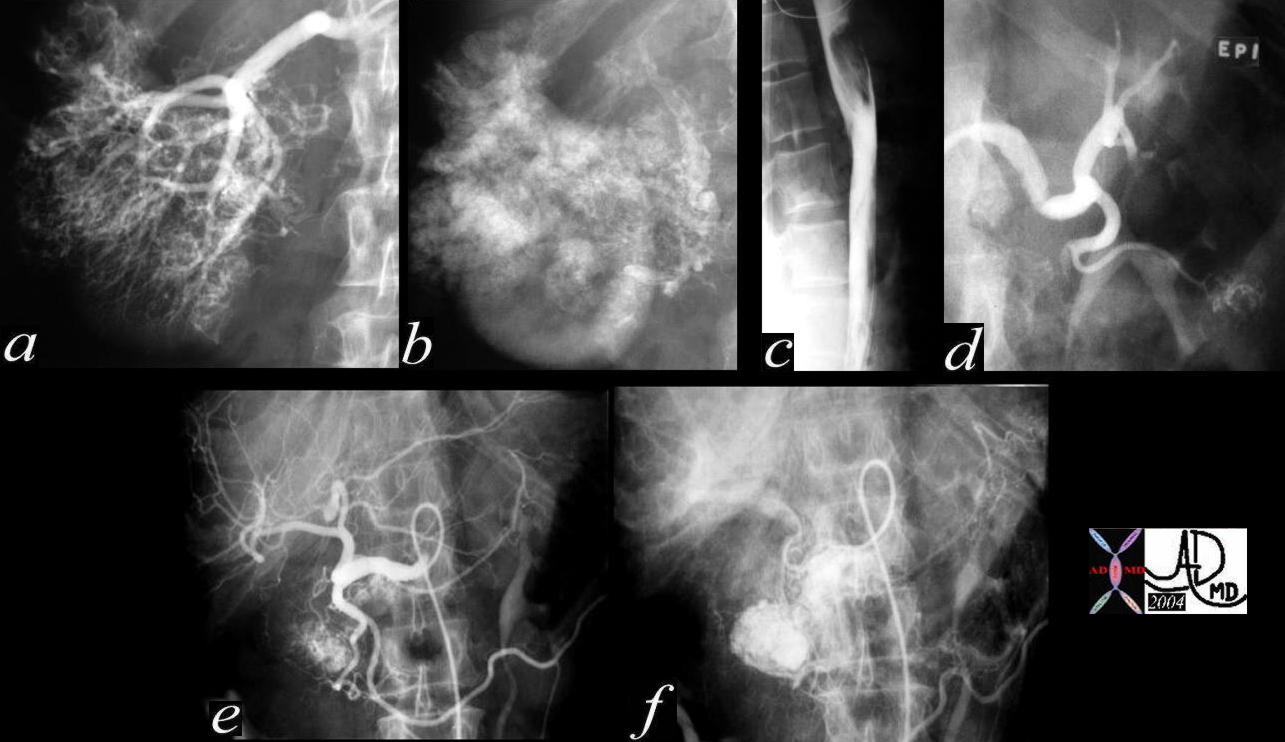
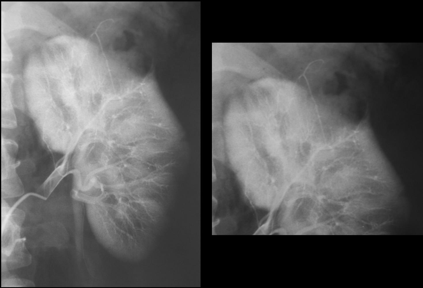
IVP and Angiography
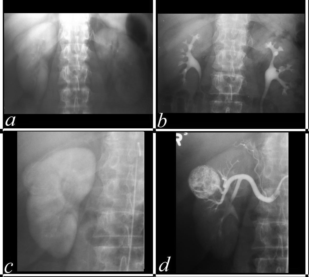
IVP and Angiogram
a IVP in the nephrographic phase shows an enhancing mass off the upper pole of the right kidney.
b – IVP in the pyelographic phase shows an a deformity of the upper and outer part of the right kidney
c – Nephrographic phase of the arteriogram confirms a hyper vascular mass
d Renal angiogram with epinephrine confirms arterial enhancement of the mass with constriction of the normal vessels and failure of the abnormal response
Ashley Davidoff MD
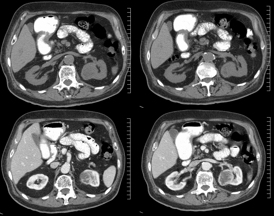
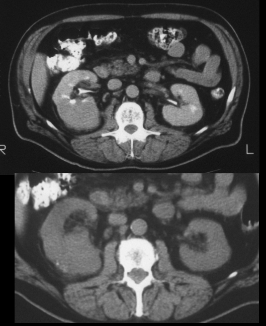
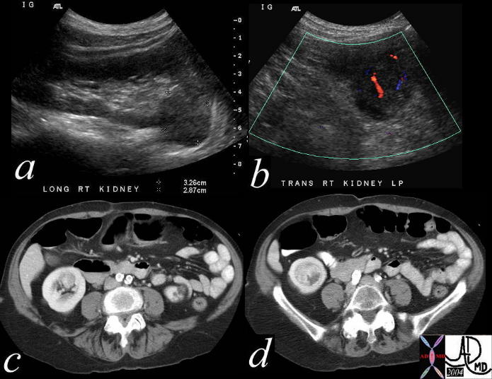
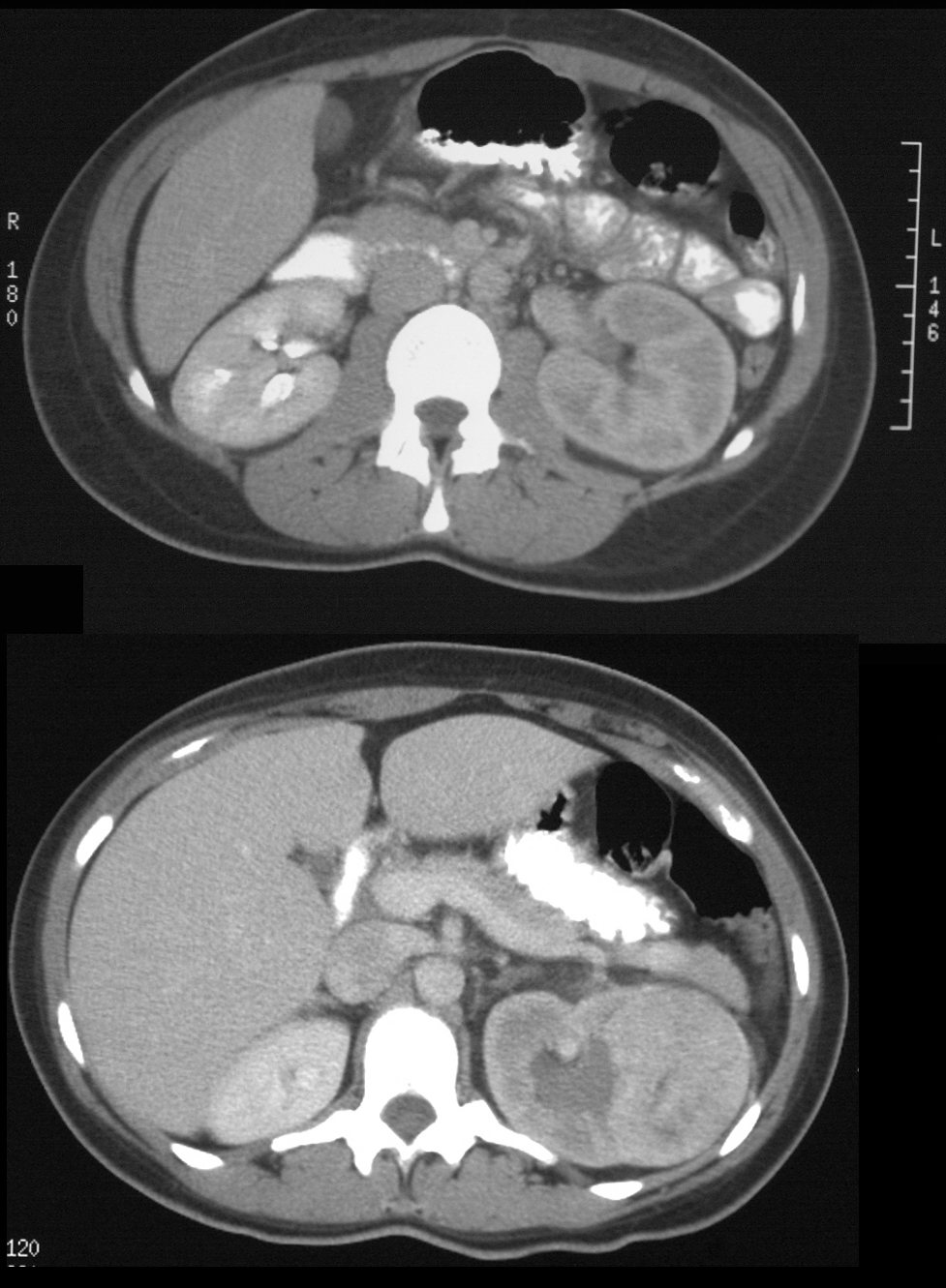
Slow Growth
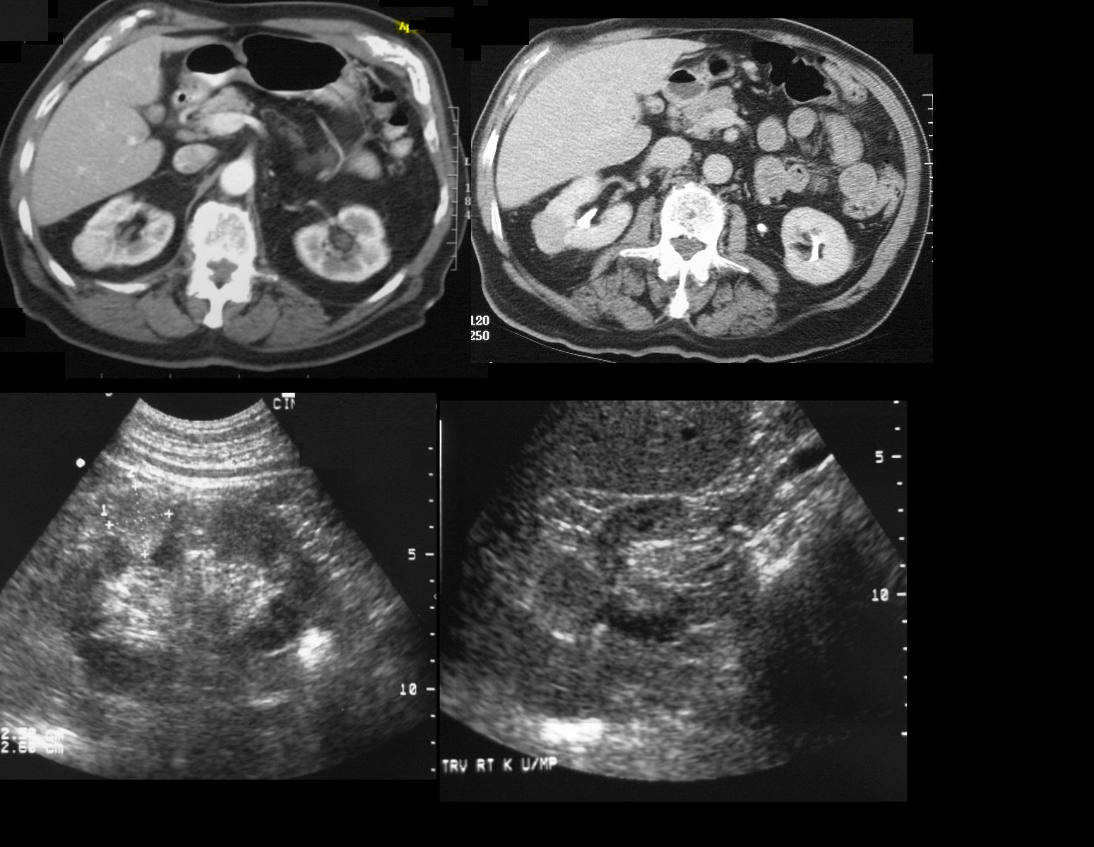
CT’ scans show two CT scans performed 2 years apart showing no significant growth
The ultrasounds below show mildly hyperechoic character of RCC
# renal cell cardcinoma#time
Ashley Davidoff MD
Ashley Davidoff MD
76776c
Invasion of the Renal Vein
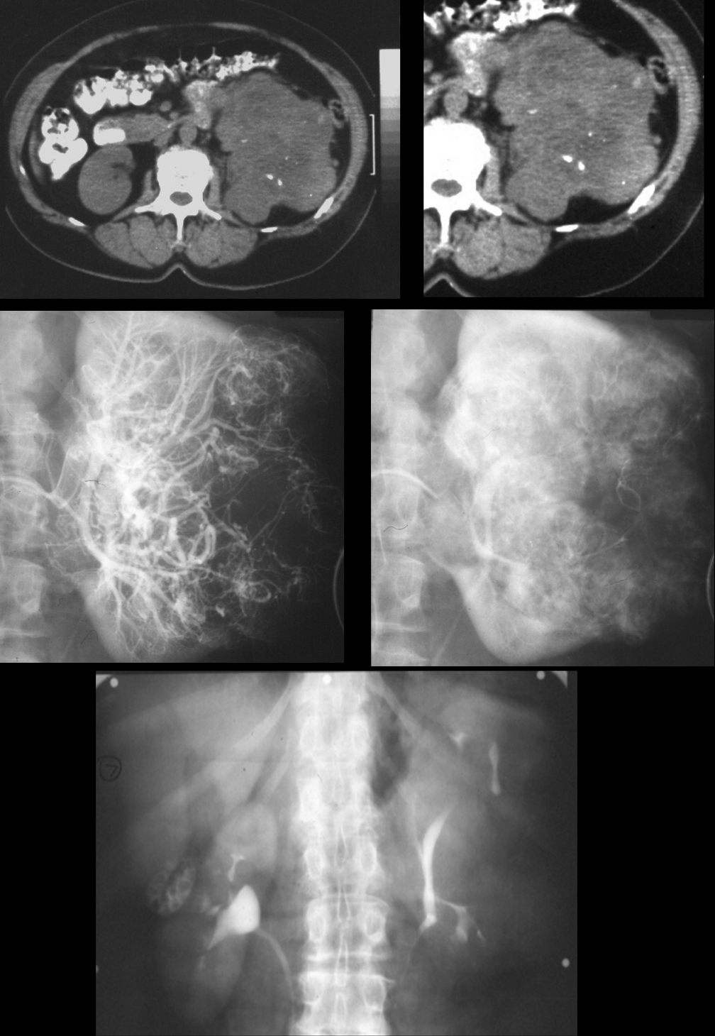
On CT there is dystrophic calcification. On angiography there is hypervascularity, neovascularity, AV shunting. Excretory phase after angiography shows distorted calyces
Ashley Davidoff MD
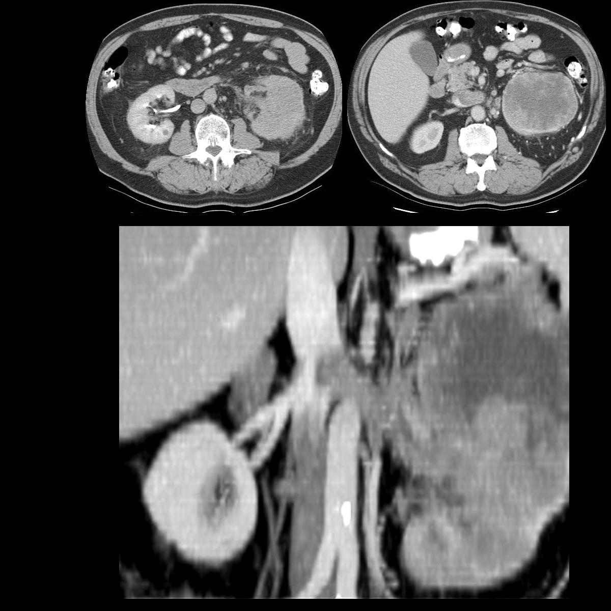
40437c Courtesy Ashley Davidoff MD code kidney renal artery fx hypervascular mass fx arteriovenous shunting fx neovascularity code IVC invasion “string and thread” sign fx filling defect code left kidney hypervascular mass code pancreas celiac axis pancreatic head fx hypervascular mass code dx primary RCC renal cell carcinoma complicated by metastases to the left kidney and pancreatic parenchyma imaging radiology angiogram venogram code neoplasm primary metastasis malignant tumor carcinoma cancer
Dystrophic CAlcification
Ashley Davidoff MD
Ashley Davidoff MD
Ashley Davidoff MD
Ashley Davidoff MD
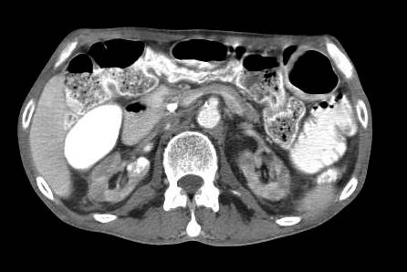
Cystic by CT
Cystic renal cell carcinoma with dystrophic calcification in the mid portion of the left kidney. Pathology reveals cystic renal cell carcinoma with clear cell histopathology
Ashley Davidoff MD
Renal Cell Carcinoma within a Cyst
Ashley Davidoff MD
34824c.1k
CT scan
Ashley Davidoff MD
Echogenic Renal Cell Carcinoma
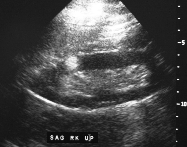
Differential consideration is an angiomyolipoma
Ashley Davidoff MD
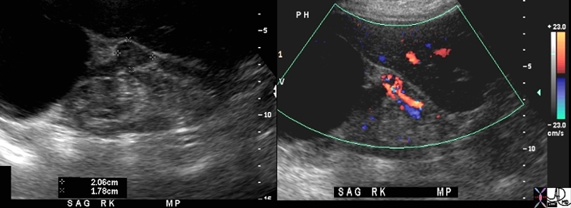
Hypervascular renal cell Carcinoma on Doppler Ultrasound
#kidney renal mass large cystic mass 2cms solid mass with hypervascularity hypervascular mass suggestion of arteriovenous shunting AV shunting large feeding arteriole and large draining vein dx renal cell carcinoma USscan Davidoff MD 48107c01
Ashley Davidoff MD
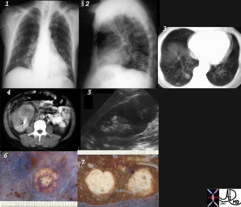
32303c01 code lungs pulmonary pleura nodules neoplasm malignant metastases metastasis primary renal kidney RCC imaging plain film CXR CTscan USscan gross pathology
Ashley Davidoff MD
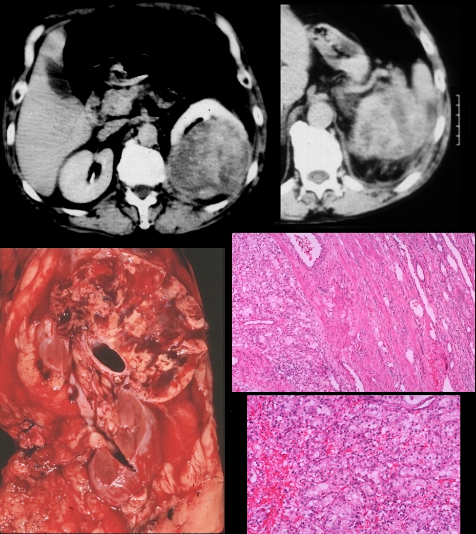
Invasion into the Right Renal Vein and IVC
Ashley Davidoff MD
Invasion into IVC and Extending to the Right Atrium (RA)
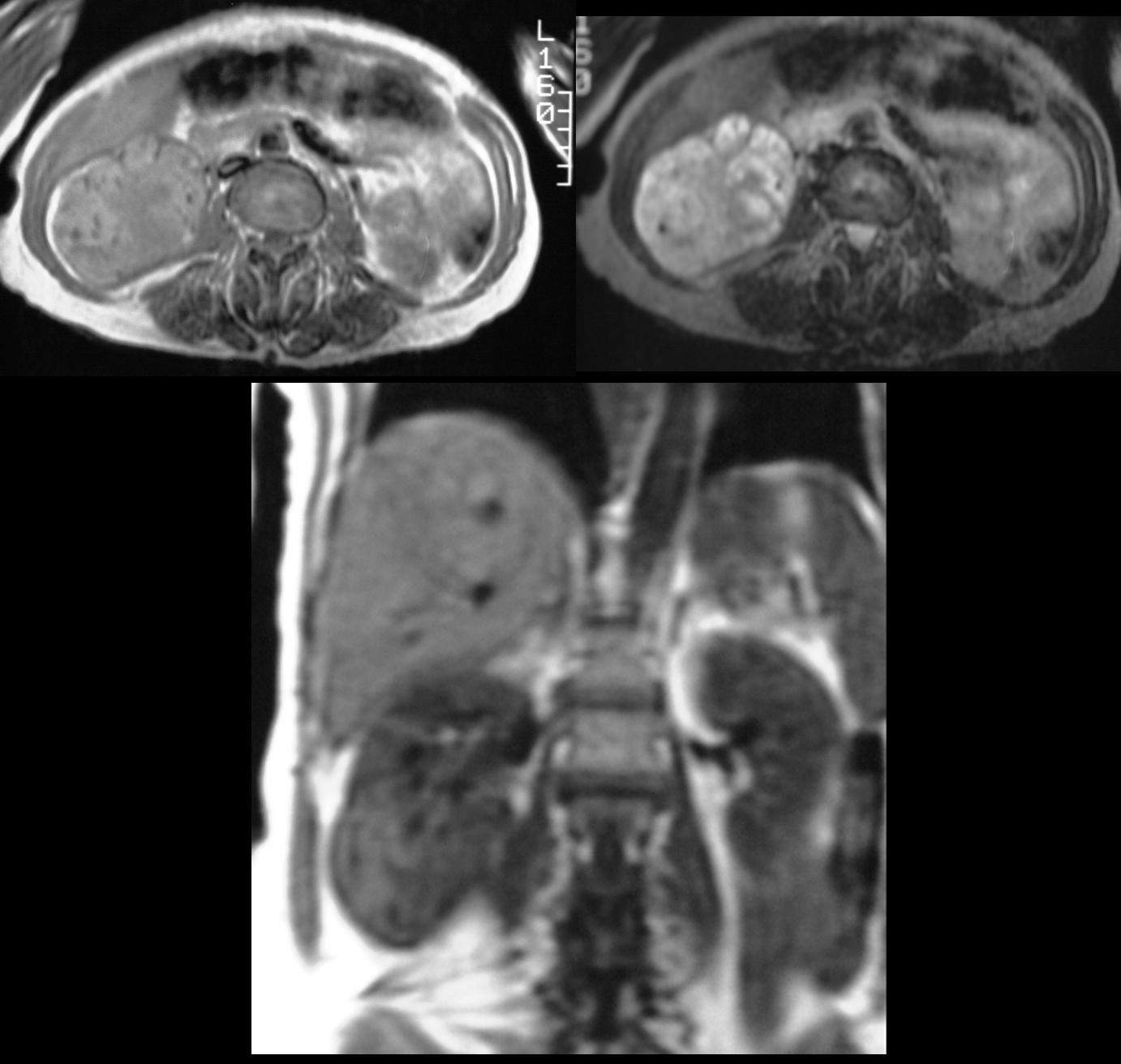
MRI (T1 and T2 ) show a large mass in the lower pole of the right kidney
Ashley Davidoff MD
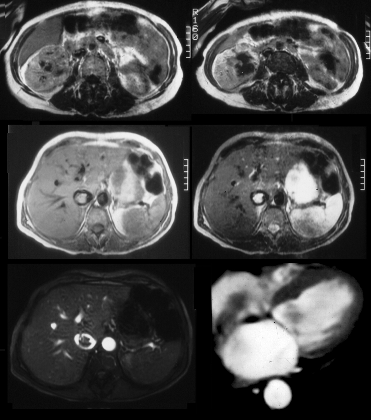
Large mass in the right kidney (T1 and T2 top row)extending into the IVC (middle row and bottom left) as well as the right atrium (RA – bottom right)
Ashley Davidoff MD

Large hypervascular mass in the right kidney . On the venous phase the filling defects with suggestion of hypervasscularity noted in the right renal vein and proximal IVC
Ashley Davidoff MD
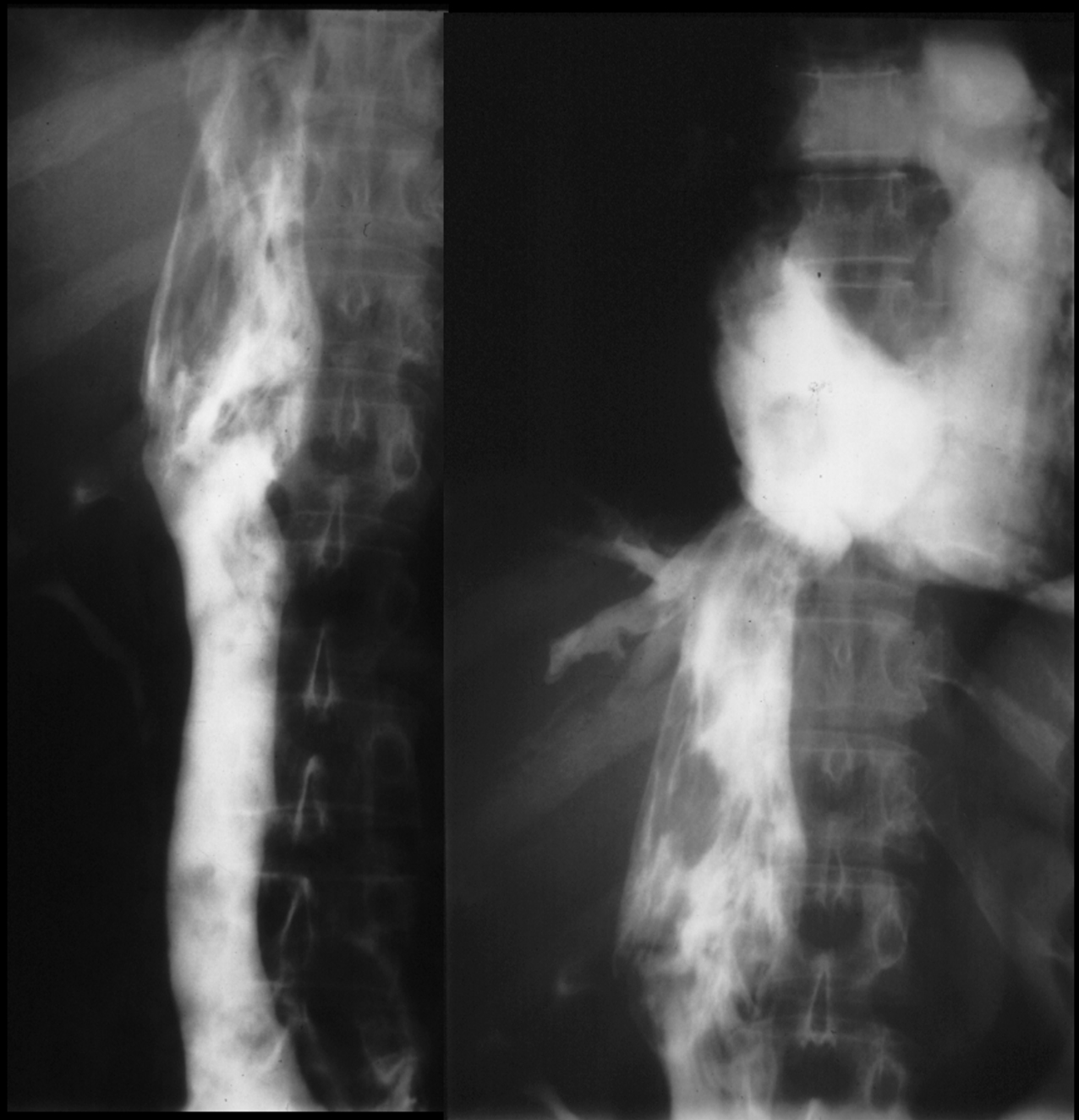
Inferior vena cavagram shows a large tumor thrombus extending into the IVC and right atrium (RA)
Ashley Davidoff MD
Renal Cell Carcinoma of the right kidney shows extension of hypervascular tumor thrombus into the right renal vein and IVC
Ashley Davidoff mD
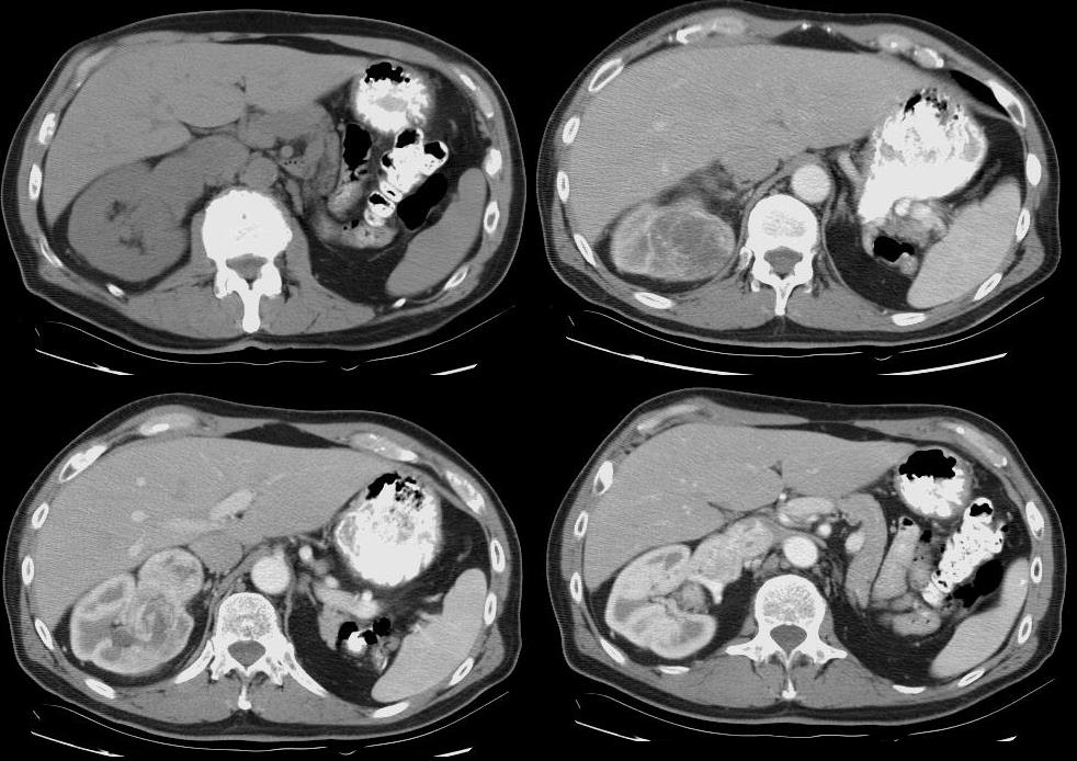
RCC Originating in the Duct of Bellini

Multiphase Contrast Enhanced MRI
Top left – Pre gadolinium shows mass like lesion
Top right Poorly visualized mass seen in the early arterial phase
Bottom 2 images – heterogeneously enhancing mass in the right upper pole
Ashley Davidoff MD
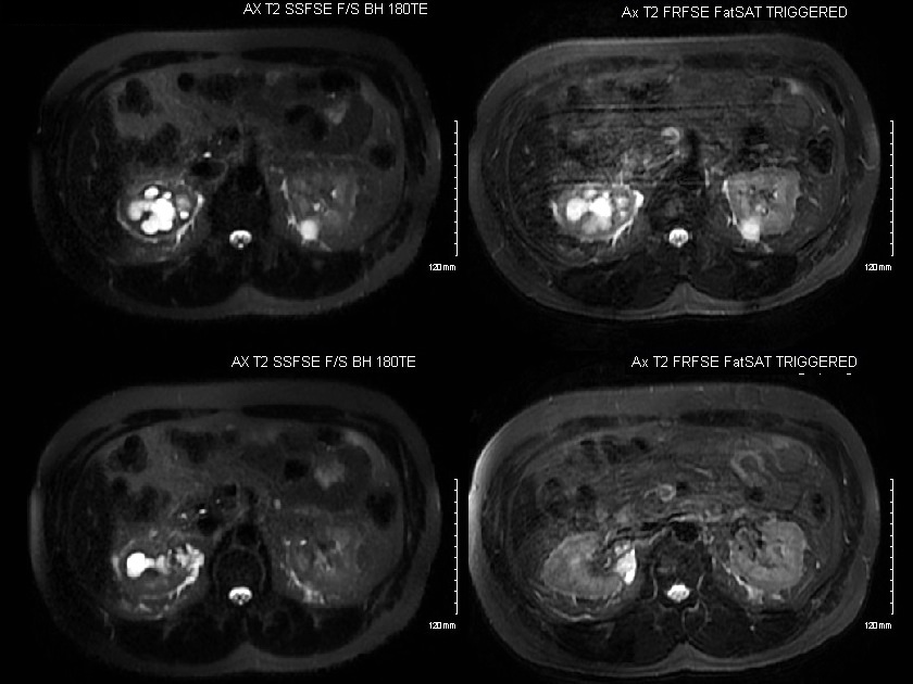
Multiphase T2 Fat Sat Images
Top right and left – Hydronephrosis right upper pole calyceal system, and mass with T2 bright component
Bottom 2 images – Perinephric fluid collection around the mass
Ashley Davidoff MD
23 F
kidney
fx delayed nephrogram
dx renal cell carcinoma
dx probable renal vein thrombosis
imaging radiology CTscan C+
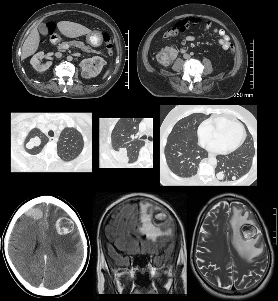
Metastases to the Lung
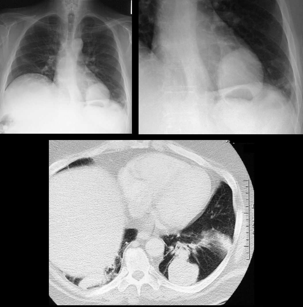
Single Large metastasis
Ashley Davidoff MD
#renal cell carcinoma
31478c
Metastases Brain
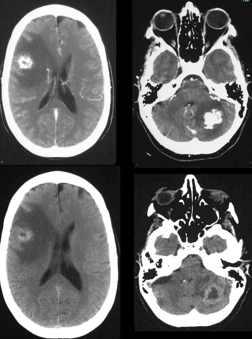
Arterial Phase (upper Images)
Delayed Phase (lower images)
brain fx mass fx hypervascular dx renal cell carcinoma dx metastasis imaging radiology CTscan C+
Ashley Davidoff MD
Ashley Davidoff MD
#renal cell carcinoma
77282c
Links and References
VF, et al Renal cell carcinoma: histological classification and correlation with imaging findings Radiol Bras vol.48 no.3 São Paulo May/June 2015