- Non Contrast Studies
-
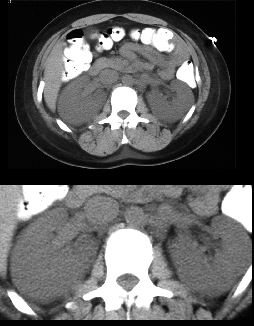
Non-Contrast CT – Acute Renal Failure and Myoglobinuria
30-year-old male presents with myoglobinuria and acute renal failure. Non-contrast CT in the axial plane in the region of the middle of the kidneys shows large, smooth, and mildly hypodense kidneys bilaterally
Ashley Davidoff MD TheCommonVein.net 16300
-
- Contrast Enhanced Studies
- Acute Tubular Necrosis
-
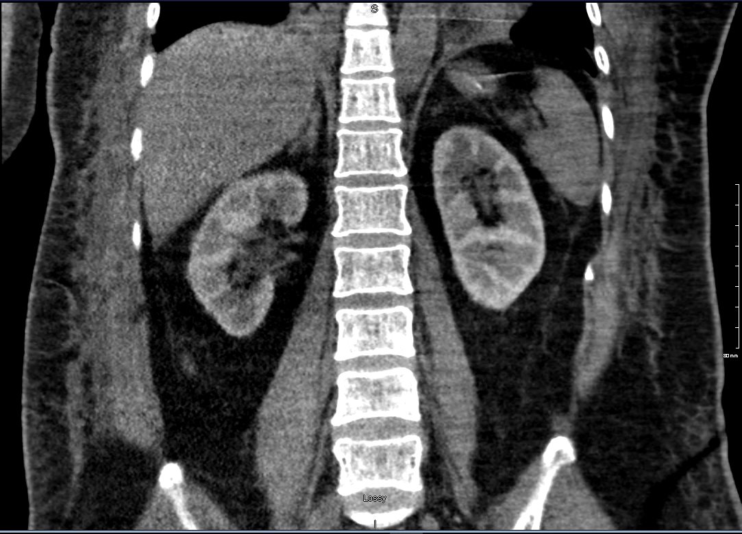
Acute Tubular Necrosis 11 days after Cardiac Catheterization
Non-contrast CT in the coronal plane in a 59-year-old female, 11 days following cardiac catheterization with acute renal failure shows persistent bilateral renal retention of contrast in the cortical phase . No contrast is seen elsewhere. Findings are consistent with acute tubular necrosis
Ashley Davidoff MD TheCommonvein.net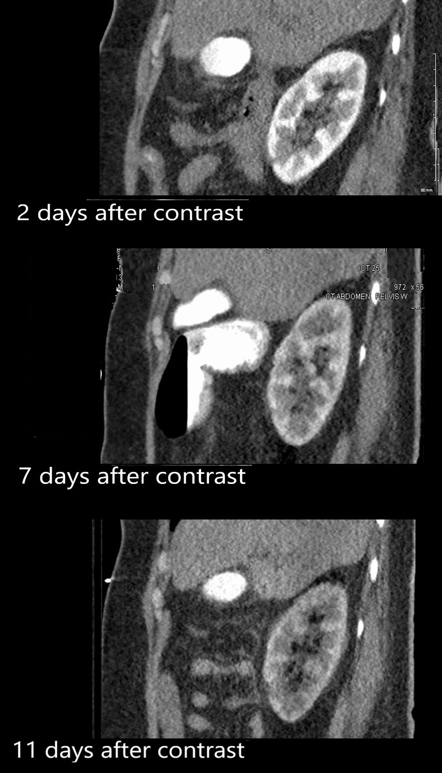
Acute Tubular Necrosis 2, 7 and 11 days after Cardiac Catheterization
Non-contrast CT in the sagittal plane in a 59-year-old female, 2, 7 and 11 days following cardiac catheterization with acute renal failure shows persistent bilateral renal retention of contrast in the cortical phase . No contrast is seen elsewhere. Findings are consistent with acute tubular necrosis
Ashley Davidoff MD TheCommonVein.net 135459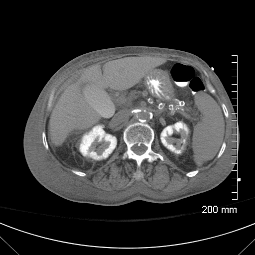
Acute Tubular Necrosis
Non-contrast CT in the axial plane in a 60 -year-old diabetic patient, following recent contrast administration now with acute renal failure. The CT shows persistent bilateral renal retention of contrast in the renal cortex . The left kidney shows regions of cortical thinning. No contrast is seen elsewhere in the vascular system. Findings are consistent with acute tubular necrosis. There is evidence of diabetic arteriopathy. The gallbladder is distended suggesting vicarious excretion. There is perinephric stranding
Ashley Davidoff MD TheCommonVein.net 132478 - Acute Cortical Necrosis
-
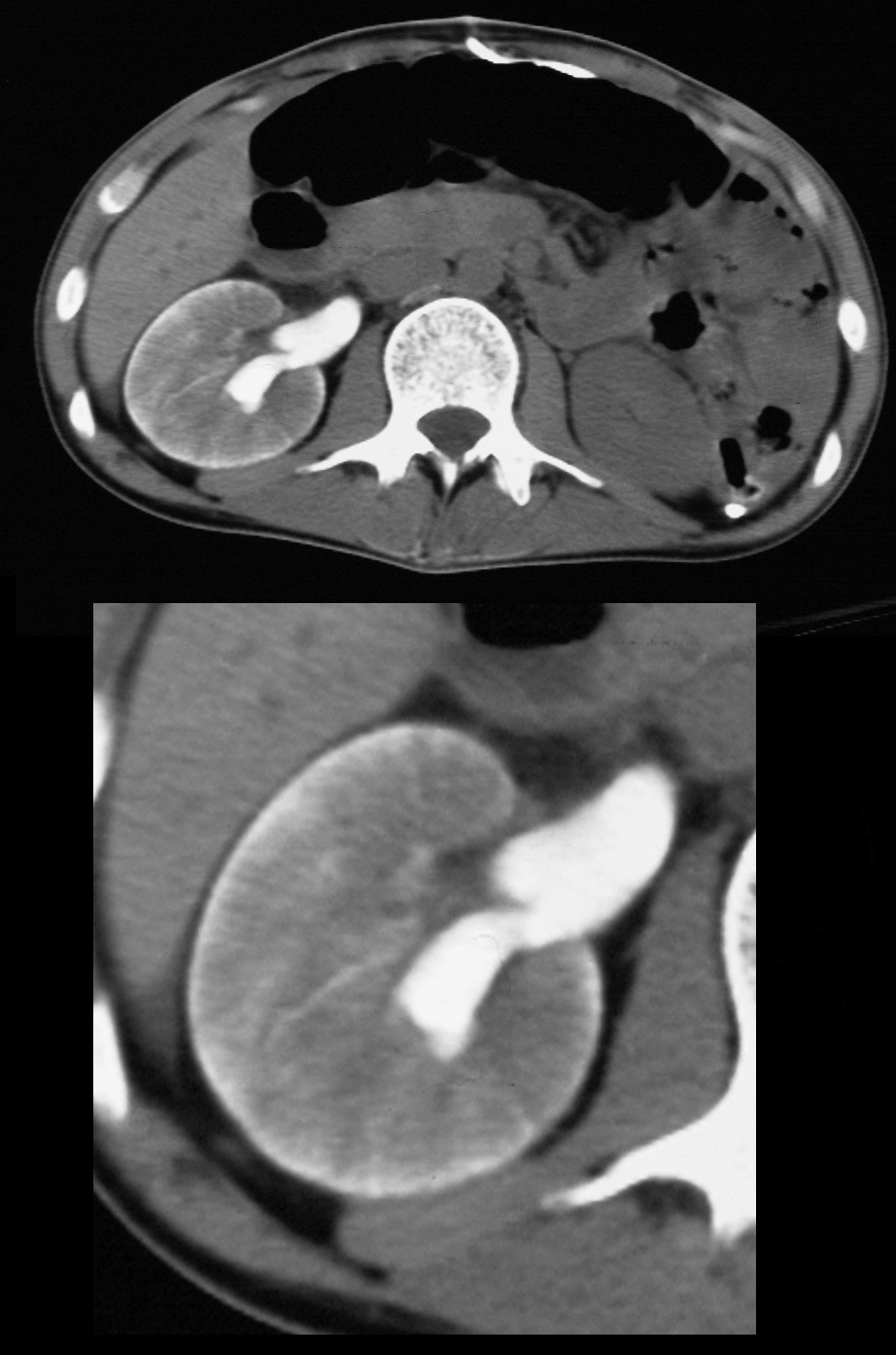
CT scan on delayed imaging shows a unilateral thin, viable rim of subcapsular cortex indicating major vascular compromise arising from acute arterial or venous compromise.
Ashley Davidoff MD TheCommonVein.net 40881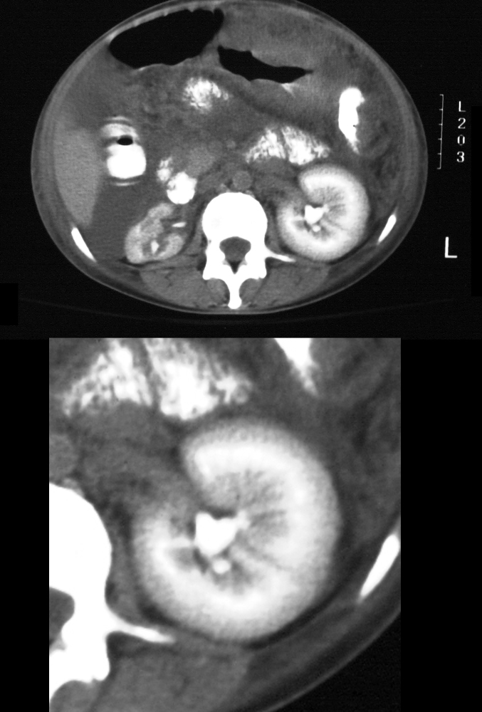
Acute Cortical Necrosis – Reverse Rim Sign
Non-contrast CT in the axial plane in a 58 -year-old patient, following contrast administration now with acute renal failure. The CT shows persistent a small right scarred kidney in the excretory phase and an enlarged left kidney with a reverse rim sign. The right kidney has a relatively low-density cortex compared to the hyper enhanced medulla. This finding known as the reversed rim sign is characteristic of acute cortical necrosis Contrast is present left renal pelvis. There is significant thickening of the stomach with ascites and suggestion of omental cake and a diagnosis of gastric cancer with omental metastases is a prime consideration
Ashley Davidoff MD TheCommonVein.net 19759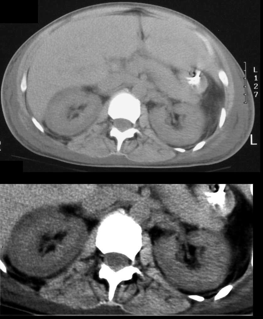
Acute Cortical Necrosis – Reverse Rim Sign
Non-contrast CT in the axial plane in a 35 -year-old female patient with microangiopathic hemolytic anemia, and acute renal failure. The non-contrast CT shows bilaterally large kidneys with a low-density rim of cortex suggesting a reverse rim sign and acute cortical necrosis. Perinephric induration is present
Ashley Davidoff MD TheCommonVein.net 20319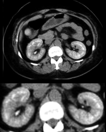
Acute Cortical Necrosis – Reverse Rim Sign
Contrast enhanced CT in the axial plane shows bilateral relatively low-density rim of cortex with a dense proximal medulla suggesting a reverse rim sign and acute cortical necrosis.
Ashley Davidoff MD TheCommonVein.net 5285
