
Inflammation Infection
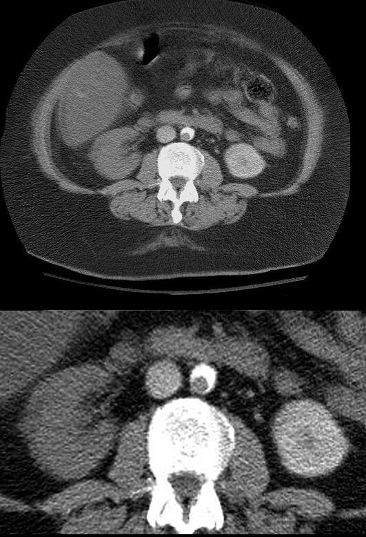
Focal Nephrographic Defect Acute Pyelonephritis CT
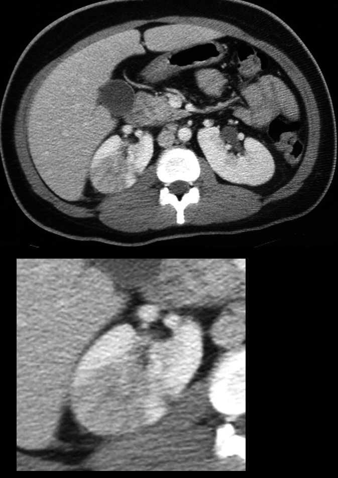
Axial CT through the kidneys shows a focal nephrographic defect in the upper pole of the right side of a horseshoe kidney. There is mils perinephric prominence of the associated posterior peritoneum. This patient presented with fever, back pain had white cells in her urine . Finding are consistent with acute pyelonephritis
Ashley Davidoff MD TheCommonVein.net 29229
Neoplasm
Trauma
Blunt Trauma, Delayed Nephrogram and Perfusion Defects Question Vascular Injury and Vasospasm
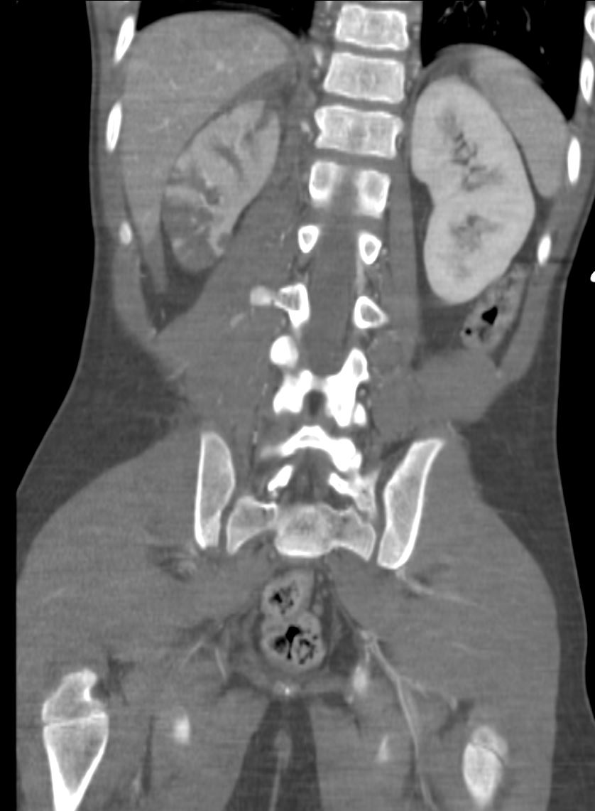
12-year-old male with hematuria and flank pain following blunt trauma to the right flank after MVA. Coronal CT through the kidneys shows delayed nephrogram and multiple wedge-shaped perfusion defects in the right kidney consistent with a vascular injury possibly related to vasospasm. There is swelling of the ipsilateral erector spinae muscle and psoas muscle.
Ashley Davidoff MD TheCommonVein.net 135572
Trauma Renal Artery Occlusion Grade V Injury
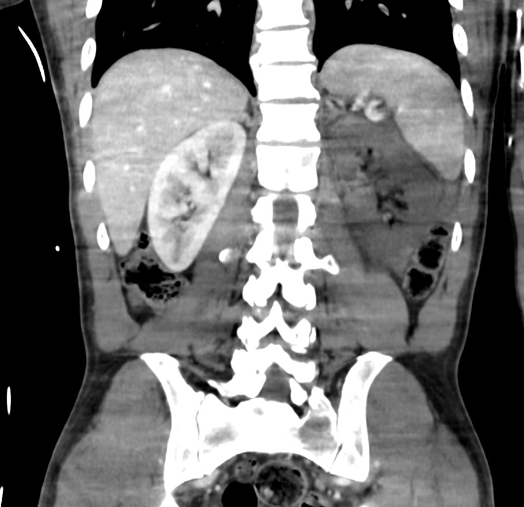
41-year-old male sustained multiple rib fractures and spine fractures in an MVA. CTA in the coronal plane shows no perfusion of the left kidney.
Ashley Davidoff MD TheCommonVein.net 135688

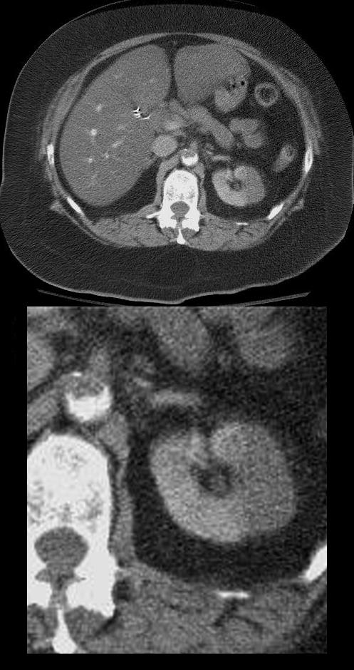
Axial CT scan in the pyelographic phase through the upper abdomen in a patient who presented with blunt trauma to the right kidney. There is a perfusion defect in the anterior aspect of the right kidney that involves both the cortex and medulla and extends to the capsular surface with evidence of thin rim of cortical enhancement, due to capsular perfusion. (“cortical rim sign”) and suggesting segmental arterial compromise.
Ashley Davidoff MD TheCommonVein.net 20267
Mechanical
Circulatory
CT Paradoxical Embolus involving the
Aorta and Left Kidney with a
Left Renal Segmental Ischemic Defect and Right Kidney Total Ischemic Perfusion Deficit
Patient presented with dyspnea and chest pain. A diagnosis of large pulmonary embolus with pulmonary infarction was made on CTPA. This image shows an embolus in the aorta and segmental non-perfusion of the left kidney as a result paradoxical embolic occlusion
Ashley Davidoff MD TheCommonVein.net 19453
CT Paradoxical Embolus Aorta and Right Renal Segmental Ischemia
Patient presented with dyspnea and chest pain. A diagnosis of large pulmonary embolus with pulmonary infarction was made on CTPA. In addition, paradoxical emboli were identified in the aorta, with non-perfusion of the right kidney and occlusion of the right iliac artery. This image shows an embolus in the aorta and non-perfusion of the right kidney as a result embolic occlusion
Ashley Davidoff MD TheCommonVein.net 19463
Blunt Trauma to the Right Kidney – Grade III Injury Segmental Ischemia – and Rim Sign
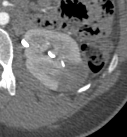
CT scan in the axial plane in a patient with flank pain shows a wedge-shaped swollen perfusion defect involving both the cortex and medulla of the left kidney and extending to the capsular surface. There is a hint of a capsular perfusion defect (cortical rim sign) .
Ashley Davidoff MD TheCommonVein.net