
Renal osteodystrophy is a group of bone changes that occur as a result of chronic kidney disease (CKD). These changes are primarily due to imbalances in calcium, phosphorus, and vitamin D metabolism, which can lead to abnormalities in bone mineralization. Imaging studies, such as X-rays and bone scans, can reveal several characteristic findings associated with renal osteodystrophy:
- Generalized Osteopenia: This refers to a decrease in bone density throughout the skeleton. On X-rays, bones may appear less dense or thinner than normal. This is a common finding in renal osteodystrophy and reflects decreased bone mineralization.
- Ruuger Jersey Spine
-
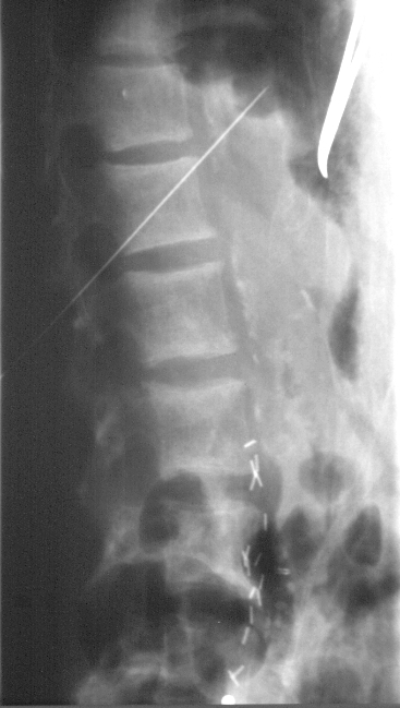
Rugger Jersey Spine in a patient undergoing translumbar aortography
Ashley Davidoff MD TheCommonVein.net #renal osteodystrophy bones lumbary spine - Osteitis Fibrosa Cystica: In more severe cases, there may be the development of bone cysts, particularly in long bones. These cysts can be seen on X-rays as lytic lesions or areas of bone resorption. They often have a ground-glass appearance.
- Subperiosteal Resorption: There may be evidence of subperiosteal resorption, especially in the phalanges. This appears as a thin, radiolucent line just below the surface of the bone.
-
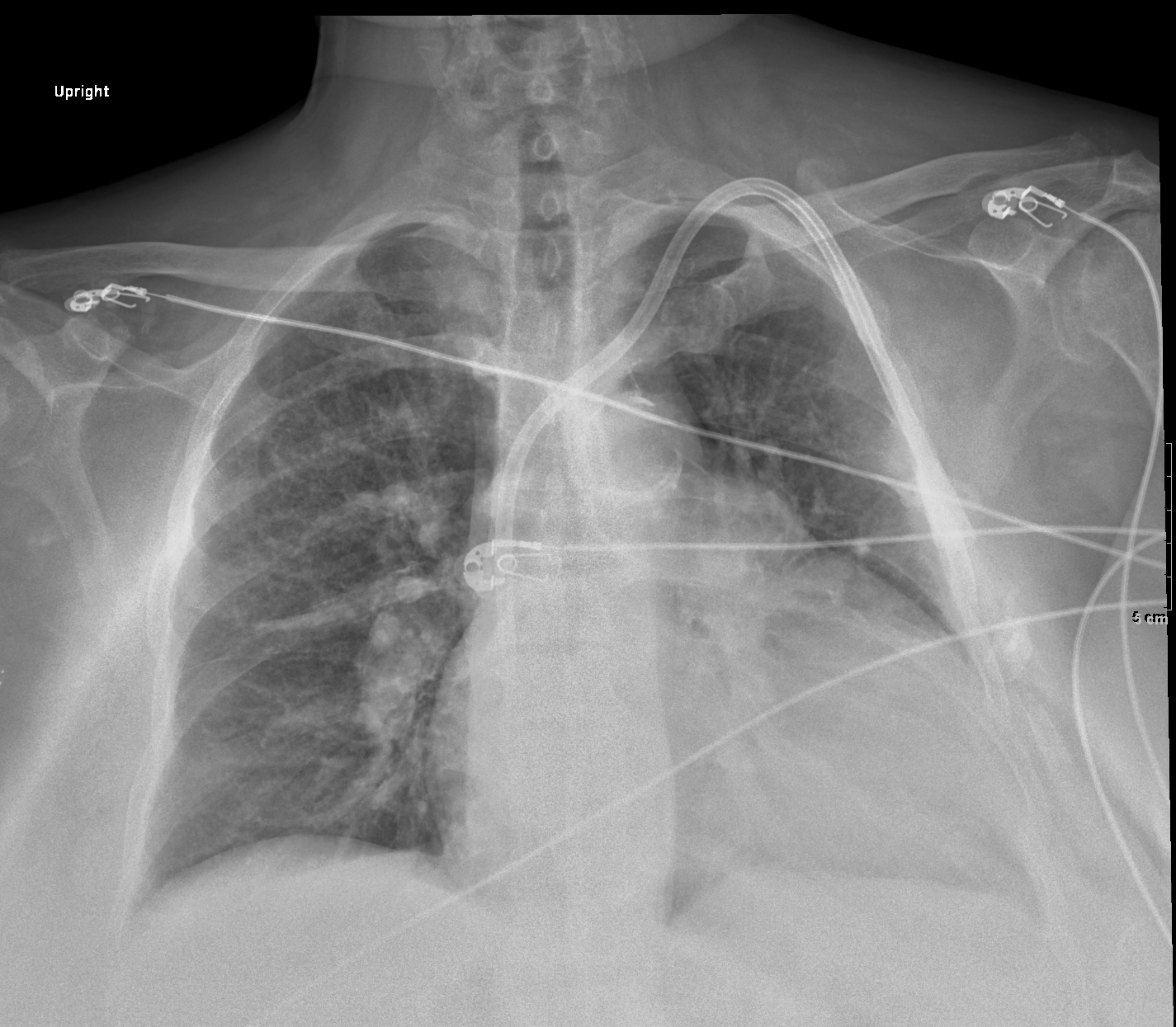
Subperiosteal Resorption of the Left Clavicle
The 68 year female has chronic renal failure (CRF) and the left clavicle shows subperiosteal resorption – a manifestation of renal osteodystrophy as a result of aberrant calcium, phosphorus, and vitamin D metabolism. Note the tunneled renal dialysis catheter used for dialysis , cardiomegaly with evidence of mild CHF
Ashley Davidoff TheCommonvein.net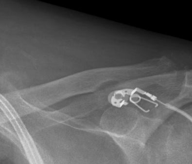
The 68 year female has chronic renal failure and the left clavicle shows subperiosteal resorption – a manifestation of renal osteodystrophy as a result of aberrant calcium, phosphorus, and vitamin D metabolism,.
Ashley Davidoff TheCommonvein.net
- Salt-and-Pepper Skull: In some cases, the skull may show a speckled or salt-and-pepper appearance on X-rays. This is due to increased bone turnover and resorption in the calvaria.
- Vascular Calcifications: Patients with renal osteodystrophy may also have vascular calcifications, which can be seen on imaging studies. These calcifications can affect blood vessels throughout the body and are often associated with cardiovascular complications.
- Fractures: Patients with renal osteodystrophy are at an increased risk of bone fractures due to the weakened bones. Fractures may be evident on X-rays, and they can occur with minimal trauma.
- Secondary Hyperparathyroidism: In cases where secondary hyperparathyroidism is present, X-rays may show evidence of soft tissue calcifications in the parathyroid glands.
- Joint Changes: Joint changes may be observed in some patients, including soft tissue calcifications around the joints or erosions of the joint surfaces.