
Class I
well-defined thin (≤2 mm) smooth wall –
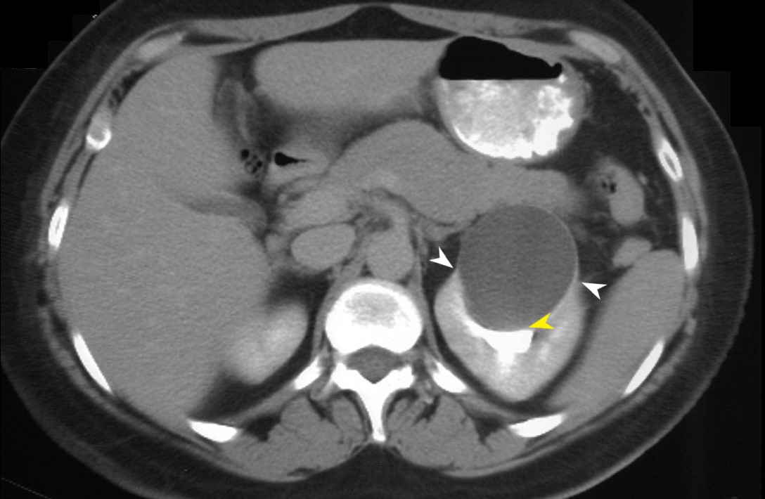
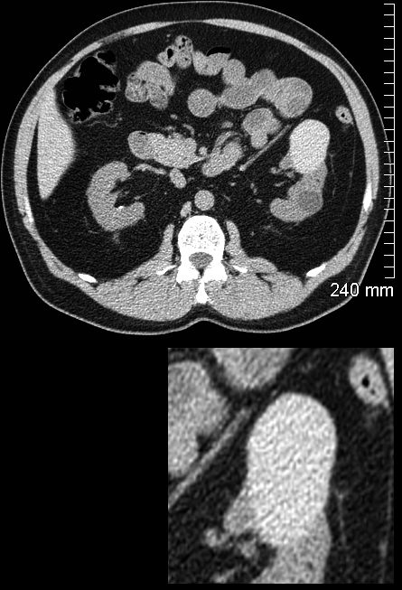
55-year-old female presents with abdominal pain. An axial CT scan through the upper portions of the left kidney shows distortion and compression of the upper pole calyces (yellow arrowhead) . There are bilateral “beak signs” (white arrowheads) indicating a benign slow growing cyst
Diagnosis: Left Sided Simple Renal Cyst Bosniak 1
Ashley Davidoff MD TheCommonVein.net RnD
Homogeneous Simple Fluid (-9 to 20 HU)
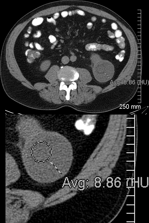
Non-Contrast CT in the axial CT plane shows a low density lesion in the left kidney with a thin wall measuring 8.9HU consistent with a cyst
Ashley Davidoff MD TheCommonVein.net 130641
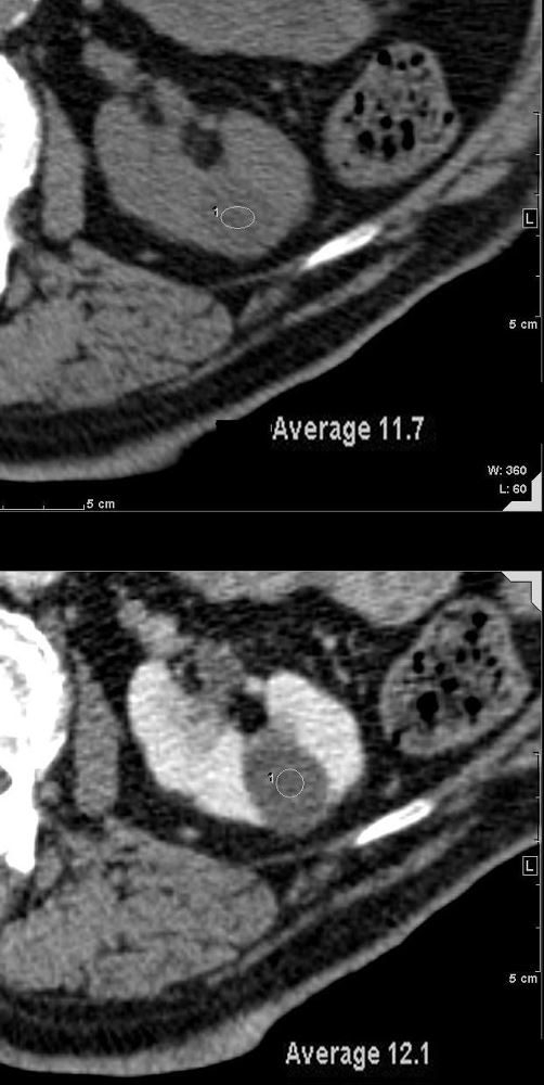
CT through the left kidney shows a low-density cyst prior to contrast measuring 11.7HU (upper image). Following contrast (lower image), the density of the nodule remained at 12 HU. The low density and lack of enhancement indicates a benign renal cyst
Ashley Davidoff MD TheCommonVein.net 135676
Class I
The Wall may Enhance after the Administration of Contrast
55-year-old female presents with abdominal pain. An axial CT scan through the upper portions of the left kidney shows distortion and compression of the upper pole calyces (yellow arrowhead) . There are bilateral “beak signs” (white arrowheads) indicating a benign slow growing cyst
Diagnosis: Left Sided Simple Renal Cyst Bosniak 1
Ashley Davidoff MD TheCommonVein.net RnD
Class II thin (≤2 mm) smooth walls
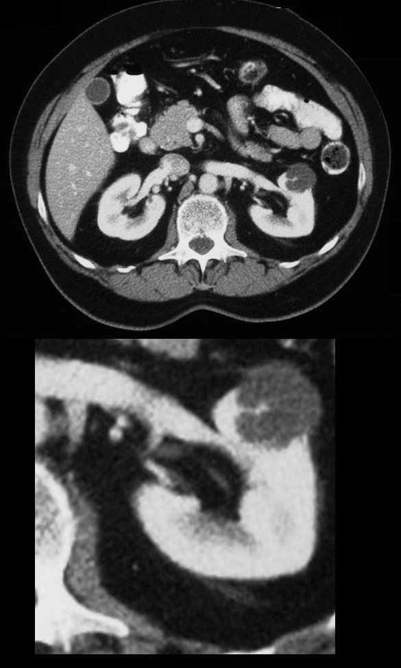
1-3 septa
CT through the mid abdomen in axial projection shows a lobular cyst with fine septations in the upper pole of the right kidney.
Ashley Davidoff MD TheCommonVein.net 130769
Class II Septa and Wall may Enhance
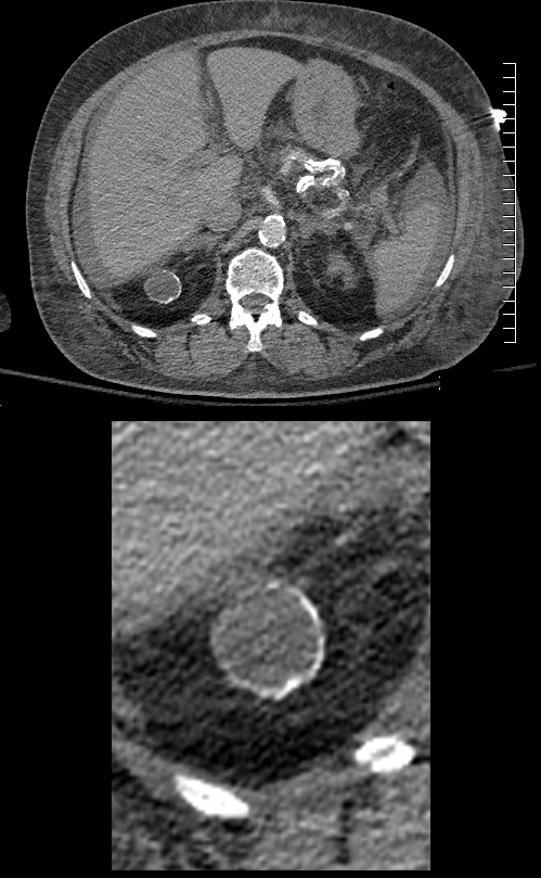
Class II Thin calcifications
CT of the upper abdomen in axial projection shows a multiple, bilateral low-density cysts. There is a fine linear calcification in the wall of a cyst in the right kidney consistent with the Bosniak 2 criterion.
Ashley Davidoff MD TheCommonVein.net 130810
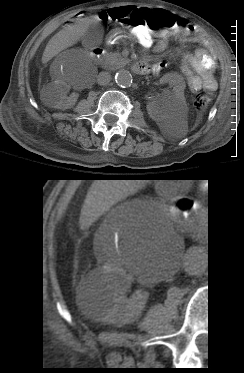
Class II F Thick or Nodular Calcification (Consider MRI)
(CT of the upper abdomen in axial projection shows a low-density cyst with coarsened calcifications in the wall. Associate findings include a small amount of ascites and subcutaneous edema in the left flank
Bosniak 2F
Ashley Davidoff MD TheCommonVein.net 135214
Class II
(≥70 HU) masses on non-contrast CT
Class II
homogeneous non-enhancing masses >20 HU at renal mass protocol CT
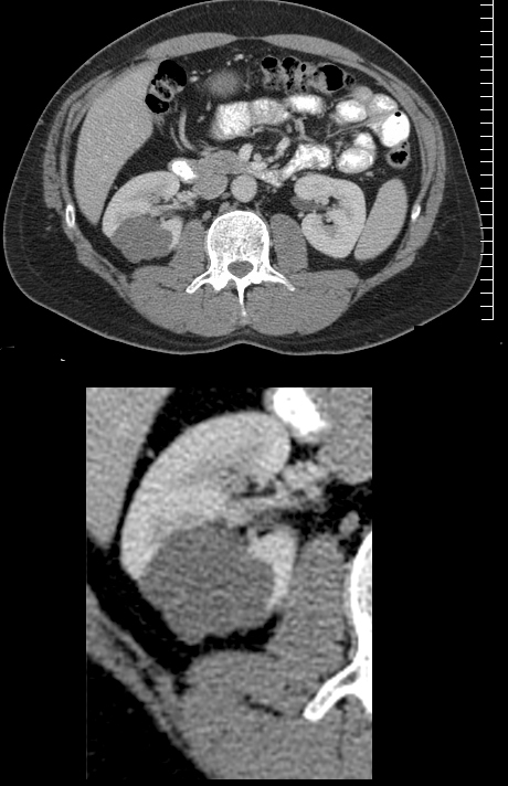
CT scan without contrast in the axial plane shows an exophytic hyperdense cyst in the mid portion of the left kidney. The commonly found simple cyst with water density is seen posteriorly for comparison. Subsequent ultrasound and MRI confirmed the benign hemorrhagic nature of the cyst
Ashley Davidoff MD TheCommonVein.net 130706
Class II
homogeneous masses (-9 to 20 HU at non-contrast CT)
homogeneous masses (21 to 30 HU at portal venous phase CT)
homogeneous low attenuation masses that are too small to characterize
Class IIF
smooth minimally thickened (3 mm) enhancing wall
smooth minimal thickening (3 mm) of one or more enhancing septa
many (≥4) smooth thin (≤2 mm) enhancing septa
Class III
one or more walls or septa that are
enhancing thick (≥4 mm width)
enhancing irregular (displaying ≤3 mm obtusely margined convex protrusion[s])
CT of the upper abdomen in axial projection shows a cyst with an enhancing septation which has a nodular component.
Ashley Davidoff MD TheCommonVein.net 30055
Class IV
one or more enhancing nodule(s)
≥4 mm convex protrusion with obtuse margins
a convex protrusion of any size that has acute margins