
38 year Old female with Complete Duplication of the right kidney and upper pole moiety showing atrophy and partial duplication of the left kidney
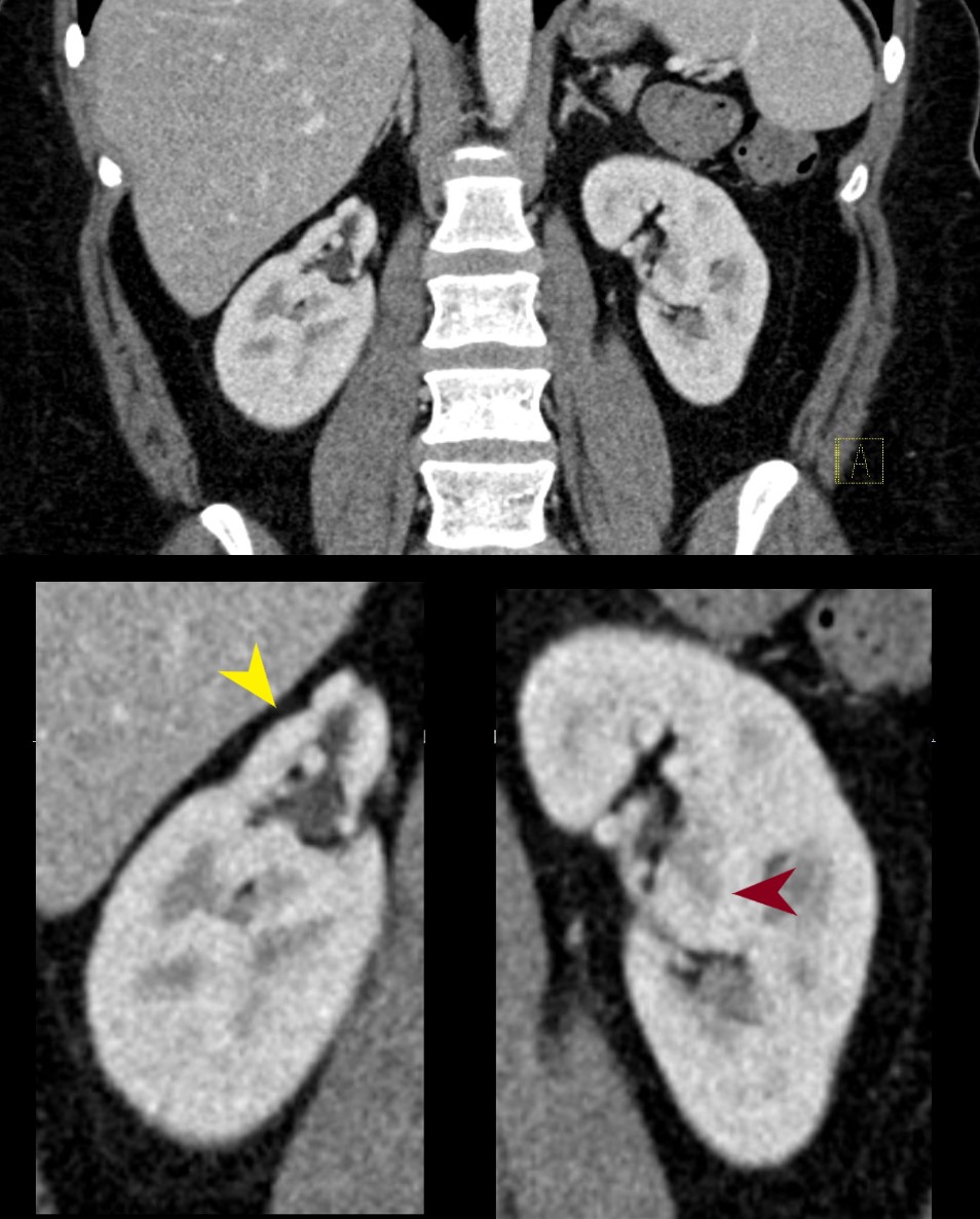
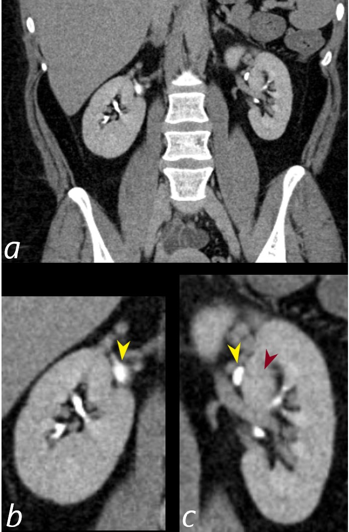
CT of the kidneys in the coronal plane in the nephrographic phase 70 seconds after injection in a 38year old female shows a duplicated right collecting system with atrophy of the upper pole moiety (yellow arrowhead lower image). The right kidney shows a prominent column of Bertin (maroon arrowhead lower image)
Ashley Davidoff MD TheCommonVein.net TCV 26K Also see 24K and 25K 135942cL
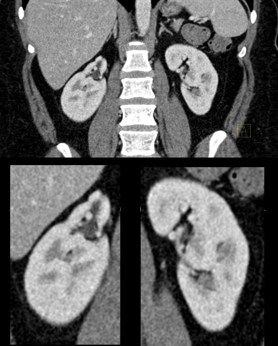
CT of the kidneys in the coronal plane in the nephrographic phase 70 seconds after injection in a 38year old female shows a duplicated right collecting system with atrophy of the upper pole moiety. The right kidney shows a prominent column of Bertin .
Ashley Davidoff MD TheCommonVein.net TCV 26K Also see 24K and 25K 135942c
SH
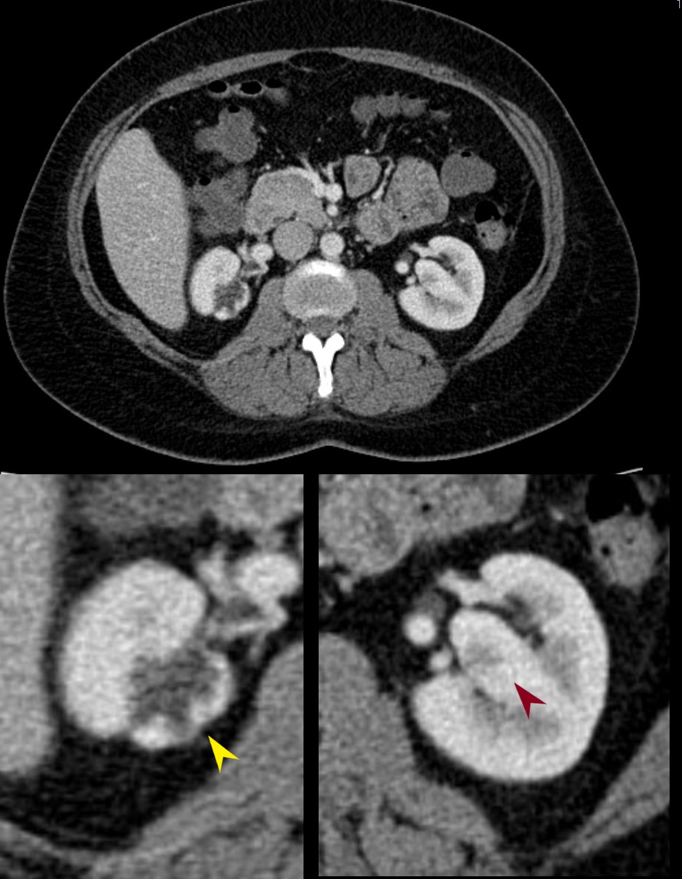
CT of the kidneys in the axial plane in the nephrographic phase 70 seconds after injection in a 38year old female shows a duplex right collecting system with atrophy of the upper pole moiety (yellow arrowhead lower image). The right kidney shows a prominent column of Bertin (maroon arrowhead lower image)
Ashley Davidoff MD TheCommonVein.net TCV 26K Also see 24K and 25K 135941cL

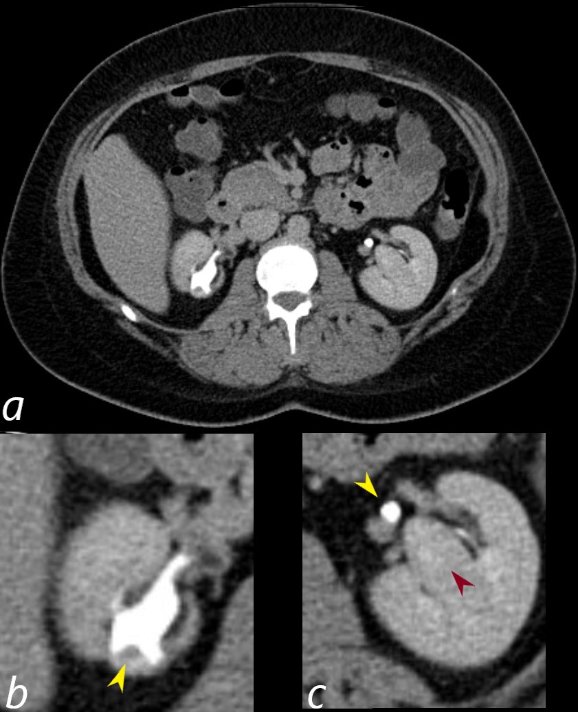
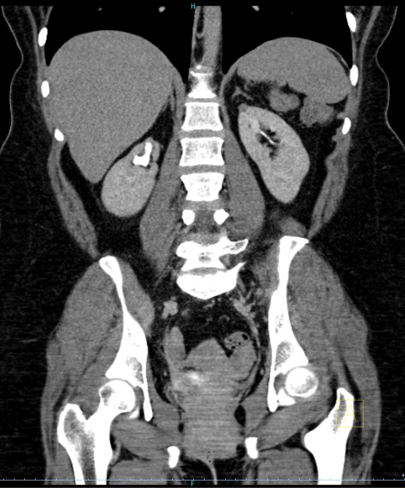
CT of the kidneys in the coronal plane in the excretory phase 10 minutes after injection in a 38year old female shows a collecting systems of the upper pole components of the duplex systems filled with contrast (yellow arrowheads b for the right side and c for the left) The column of Bertin on the left (c maroon arrowhead) separates the upper and lower moieties of the partial duplication on the left .
Ashley Davidoff MD TheCommonVein.net TCV 26K Also see 24K and 25K 135943cL
CT of the kidneys in the axial plane in the excretory phase 10 minutes after injection in a 38year old female shows a hydronephrotic collecting systems of the upper pole components of the duplex systems filled with contrast (yellow arrowheads b) and evidence of post obstructive atrophy. The left extrarenal collecting system is of normal caliber as a common ureter (c yellow arrowhead) The column of Bertin on the left is noted (c maroon arrowhead
Ashley Davidoff MD TheCommonVein.net TCV 26K Also see 24K and 25K 135943cL
CT of the kidneys in the coronal plane in the excretory phase 10 minutes after injection in a 38year old female shows a collecting systems of the upper pole components of the duplex systems filled with contrast (yellow arrowheads b for the right side and c for the left) The column of Bertin on the left (c maroon arrowhead) separates the upper and lower moieties of the partial duplication on the left .
Ashley Davidoff MD TheCommonVein.net TCV 26K Also see 24K and 25K 135943cL
CT of the kidneys in the coronal plane in the excretory phase 10 minutes after injection in a 38year old female shows a hydronephrotic collecting systems of the upper pole components of the duplex systems filled with contrast with post obstructive atrophy
Ashley Davidoff MD TheCommonVein.net TCV 26K Also see 24K and 25K 135948
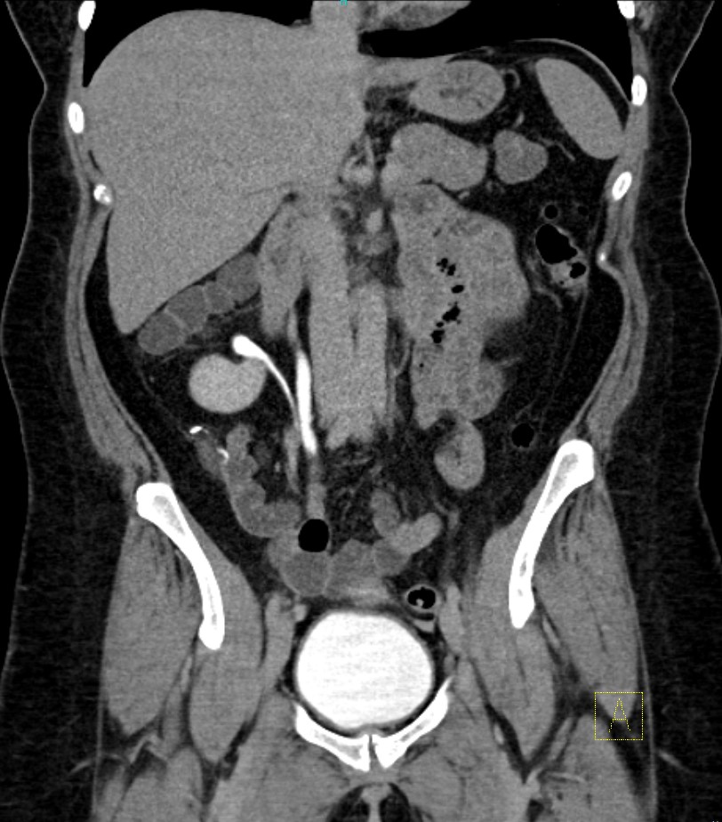
CT of the kidneys in the coronal plane in the excretory phase 10 minutes after injection in a 38year old female shows mild hydroureter of the upper pole moiety (medial ureter) and normal caliber ureter of the right lower moiety (latera ureter)
Ashley Davidoff MD TheCommonVein.net TCV 26K Also see 24K and 25K 135950
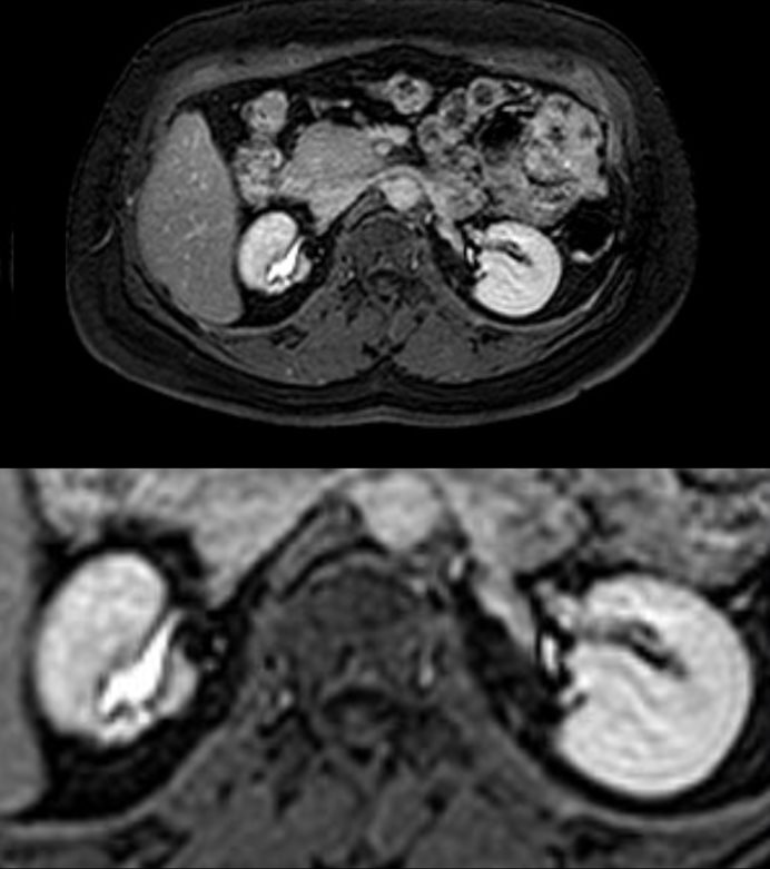
MRI Pre Contrast
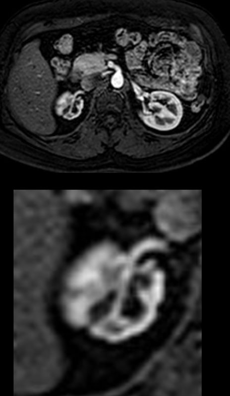
Pre contrast T1 weighted MRI through the kidneys show the atrophied right upper pole moiety in a 38year old female
Ashley Davidoff MD TheCommonVein.net TCV 25K Also see 24K and 26K 135951c
Cortical Phase at 20 Seconds after Contrast
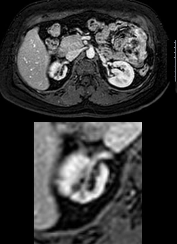
20 seconds following contrast, the T1 weighted MRI through the kidneys show the atrophy of the right upper pole moiety with irregular thinning of the right renal cortex
Ashley Davidoff MD TheCommonVein.net TCV 25K Also see 24K and 26K 135952c
Corticomedullary Phase at 70 Seconds
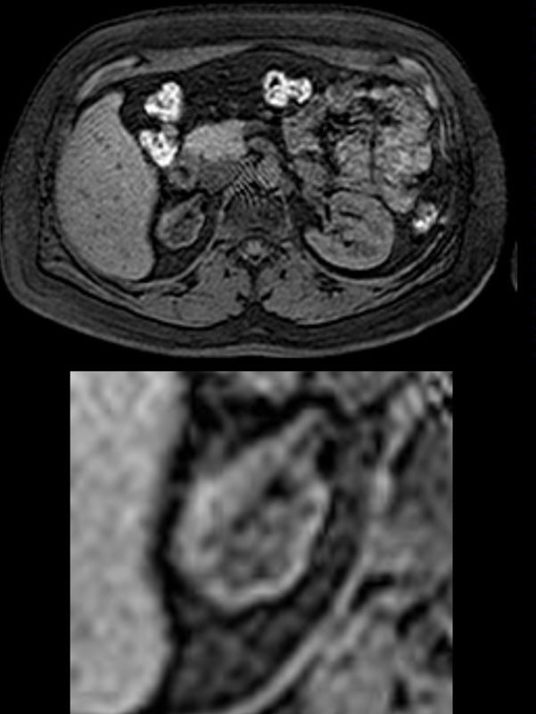
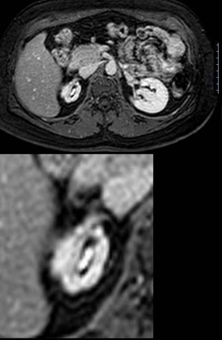
70 seconds following contrast, the T1 weighted MRI through the kidneys show the atrophy of the right upper pole moiety with indication of perfusion of the atrophied corticomedullary parenchyma. The right lateral edge of the atrophied kidney does partial volume with the normal lower pole parenchyma, and therefore the medial parenchyma reflects the true parenchyma of the upper pole moiety. Note also the column of Bertin of the left kidney
Ashley Davidoff MD TheCommonVein.net TCV 25K Also see 24K and 26K 135956c
Nephrographic and Excretory Phase at 3 minutes after Contrast –
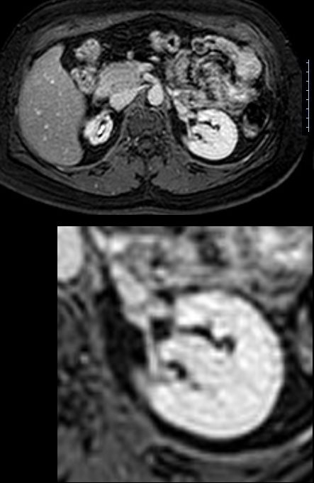
3 minutes following contrast, the T1 weighted MRI through the kidneys show atrophy of the right upper pole moiety with indication of progressive perfusion of the atrophied parenchyma. The right lateral edge of the atrophied kidney does partial volume with the normal lower pole parenchyma, and therefore the medial parenchyma reflects the true parenchyma of the upper pole moiety. Contrast is now also seen in the collecting system of the right kidney. A column of Bertin is noted in the left kidney
Ashley Davidoff MD TheCommonVein.net TCV 25K Also see 24K and 26K 135954c
Ashley Davidoff MD TheCommonVein.net TCV 25K Also see 24K and 26K 135954c01
The dilated intrarenal collecting system of the upper pole moiety is seen with associated pressure atrophy. A column of Bertin is noted extending toward the hilum of the left kidney
Ashley Davidoff MD TheCommonVein.net TCV 25K Also see 24K and 26K 135955