
CT Chronic Renal Failure Persistent Nephrogram
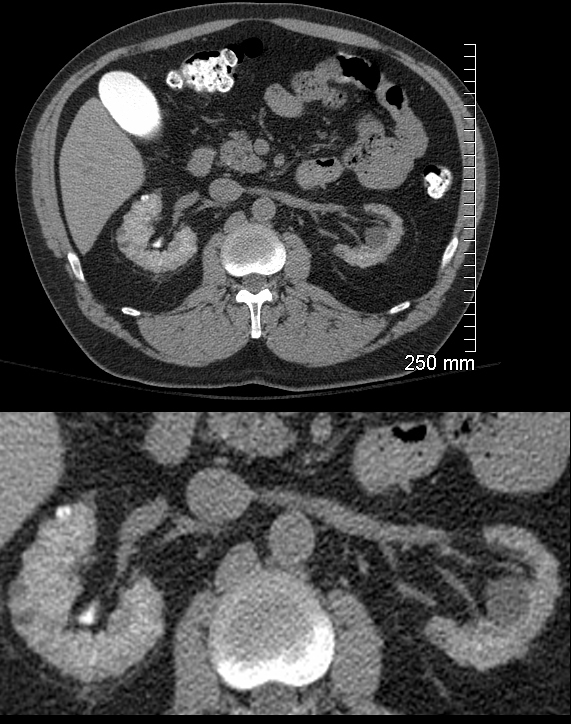
CT in the axial plane through the kidneys shows bilateral small irregular kidneys, left smaller than right with persistent nephrograms a day after a cardiac catheterization. While the larger right kidney does show some excretion into the calyces, there is no identifiable excretory phase in the left kidney. Neither the aorta nor the IVC has contrast and there is vicarious excretion of contrast in the gallbladder which is further evidence of chronic renal failure.
Ashley Davidoff MD TheCommonVein.net 130983c
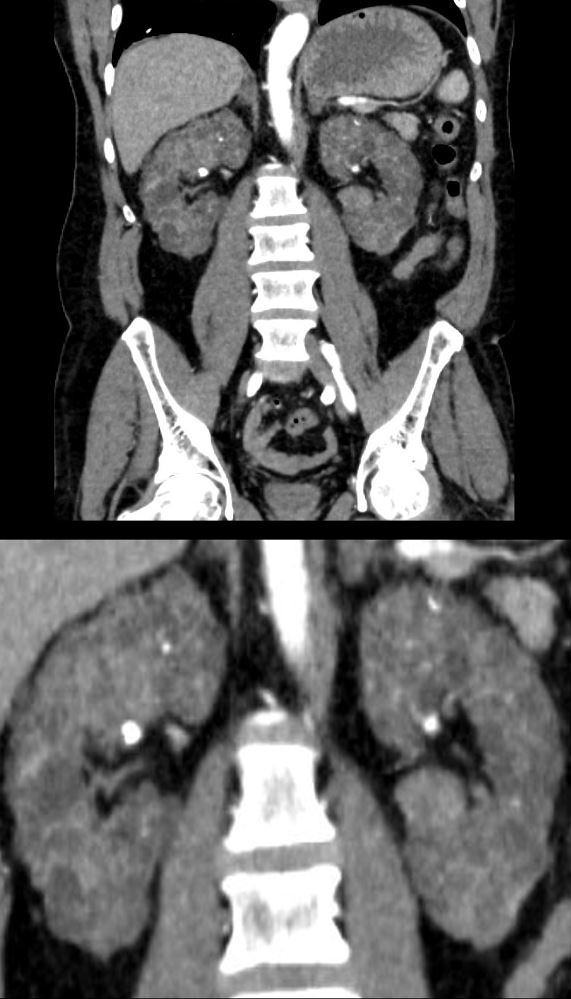
Thin Irregular Enhancing Cortex and Renal Sinus Lipomatosis
Ashley Davidoff MD TheCommonVein.net
Bilaterally Small Smooth Kidneys with Renal Sinus Lipomatosis
keywords renal atrophy small kidneys renal sinus lipomatosis Ashley Davidoff TheCommonVein.net
Ashley Davidoff MD The CommoVein.net
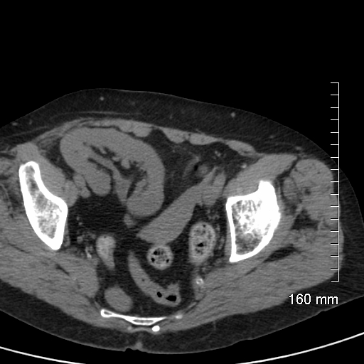
Polycystic Kidney Disease PCKD
56-year-old male in chronic renal failure presents with flank pain. CT with contrast shows diffuse cystic changes of near normal sized kidneys with total replacement of the renal parenchyma by small uniformly sized cysts. Multiple calcifications are scattered throughout the parenchyma.
Ashley Davidoff MD TheCommonVein.net 135723c
Ashley Davidoff MD The CommonVein.net
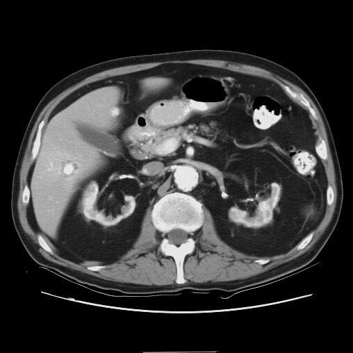
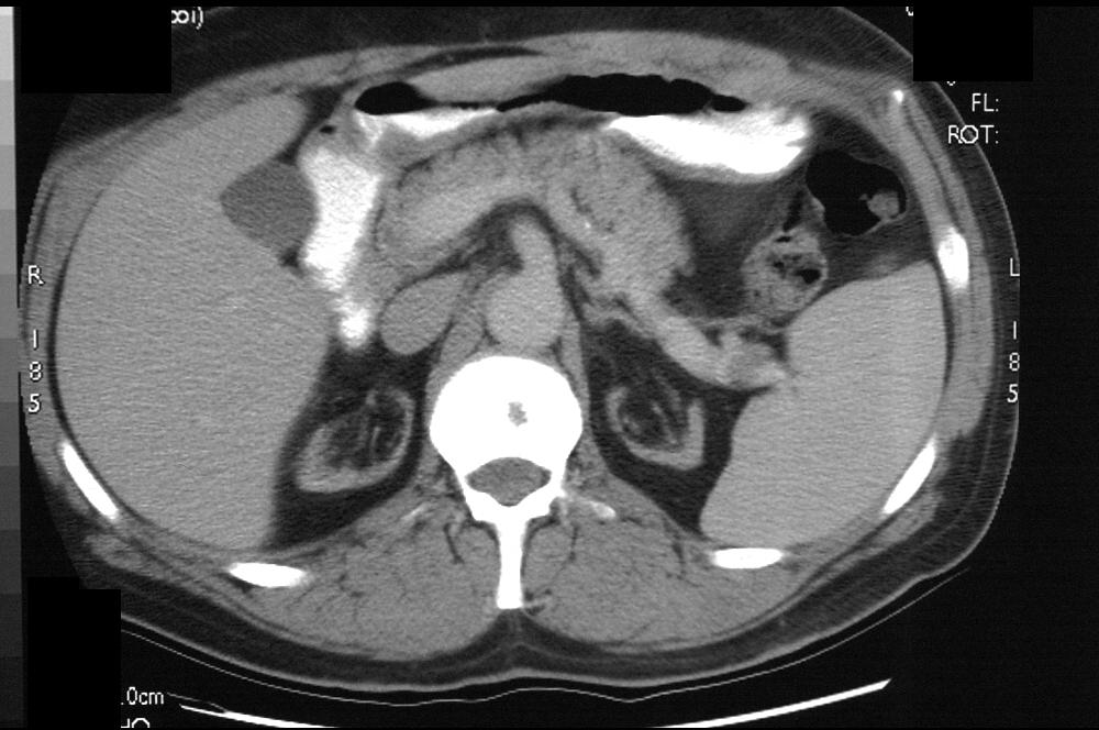
Acquired Cystic Disease of Chronic Renal Failure
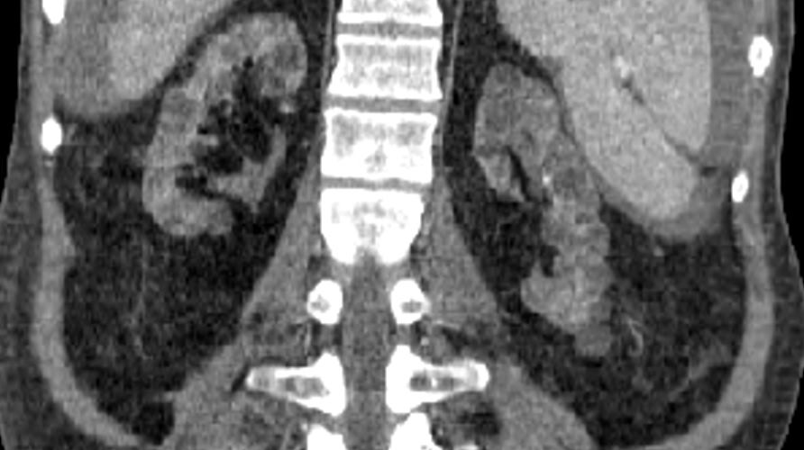
67 year old male with chronic renal failure. Reconstruction of a CT in the coronal plane shows bilateral diffuse small cysts consistent with the diagnosis of acquired cystic disease of chronic renal failure. Although the craniocaudal span is only mildly reduces there is significant parenchymal thinning and renal sinus lipomatosis. Noted ascites from peritoneal dialysis
Ashley Davidoff MD TheCommonVein.net
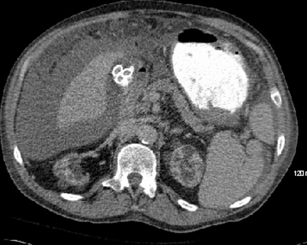
67 year old male with chronic renal failure. CT in the axial plane shows bilateral diffuse small cysts consistent with the diagnosis of acquired cystic disease of chronic renal failure. Noted ascites from peritoneal dialysis as well as induration of the greater omentum. Calcified gallstones are present
Ashley Davidoff MD TheCommonVein.net
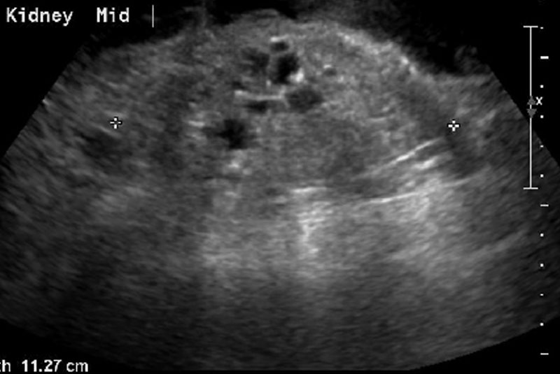
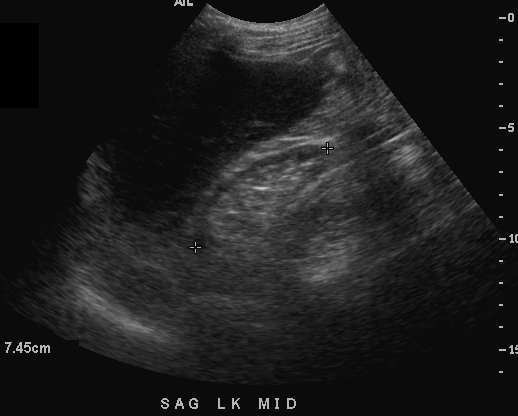
67 year old male with chronic renal failure. US in the longitudinal plane shows an 11.3cms echogenic kidney with multiple cysts in the 8mm to 10mms range consistent with the diagnosis of acquired cystic disease of chronic renal failure.
Ashley Davidoff MD TheCommonVein.net