
CT Papillary Necrosis

40-year-old female with SLE and NSAID use presents with hematuria. CT with contrast in the axial plane during the excretory shows evidence of a filling defect in a contrast filled right renal calyx, resulting in a “lobster claw” appearance of the calyx characteristic of papillary necrosis
Ashley Davidoff MD TheCommonVein.net 113597c
CT Filling Defect in a Calyx Phantom Calyx Sign
77-year-old male with smoking history presents with hematuria. CT with contrast in the axial plane during the excretory shows evidence of an expansile filling defect occupying a left upper pole calyx. Histology showed a transitional cell carcinoma (TCC), The associated imaging sign is called a ‘phantom calyx sign” inferring a calyx that completely fails to opacify
Ashley Davidoff MD TheCommonVein.net 06104c
Nephrostogram – Multiple Calyceal Filling Defects Candida
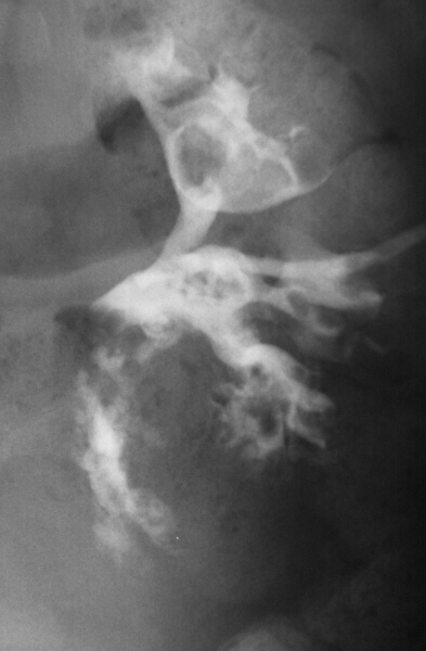
45-year-old male diabetic presents with flank pain and fever. Nephrostogram shows multiple filling defects in the calyces and throughout the intrarenal collecting system and proximal ureter. Culture of the urine revealed extensive growth of candida
Ashley Davidoff MD TheCommonVein.net 15822