
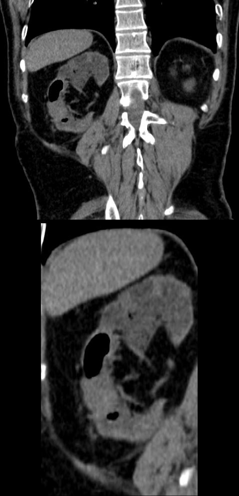
Coronal CT– Emphysematous Pyelonephritis
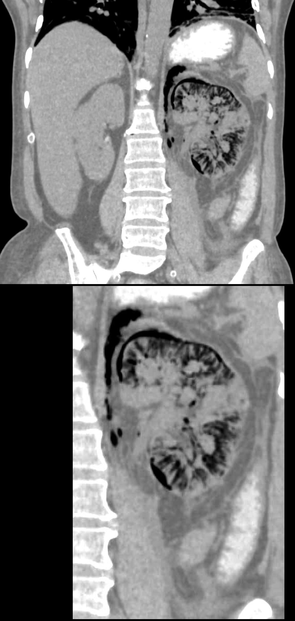
55-year-old diabetic female presents with flank pain and fever. CT without shows diffuse infiltration of air into the swollen left kidney including the intertstitium of the renal parenchyma, subcapsular regions, perinephric space and the retroperitoneum. There is perinephric induration and thickening of Gerota’s fascia and perinephric edema. Small islands of tissue are noted in the upper medial portions of the kidney and the mid portion
Ashley Davidoff MD TheCommonVein.net 135714
Emphysematous pyelonephritis (EPN) is a severe, necrotizing infection of the kidney that is characterized by the presence of gas within the renal parenchyma, collecting system, or perinephric tissues. It is typically associated with urinary tract infections, most commonly caused by bacteria such as Escherichia coli. The condition is more common in individuals with diabetes mellitus, urinary tract obstruction, or other conditions that compromise the immune system.
EPN is a medical emergency, and prompt diagnosis and treatment are essential. The presence of gas within the kidney can be visualized on imaging studies such as CT scans. The infection can spread rapidly and may lead to serious complications, including sepsis and organ failure.
Treatment usually involves a combination of antibiotics and surgical intervention. Antibiotics are administered to control the infection, and surgery may be necessary to drain the infected fluid and remove necrotic tissue. In some cases, a nephrectomy) may be required.
Due to the severity of emphysematous pyelonephritis, close monitoring in an intensive care setting may be necessary. The choice of antibiotics is guided by the culture and sensitivity results, and adjustments may be made based on the patient’s response to treatment.
Coronal CT with Contrast– Emphysematous Pyelonephritis
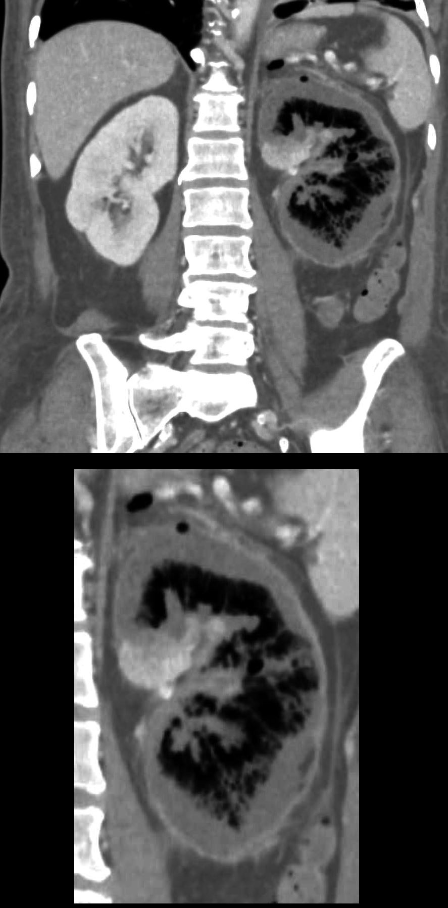
55-year-old diabetic female presents with flank pain and fever. CT with contrast in the nephrogram phase shows a non-functioning rim of cortex and medulla, and a small island of functioning parenchyma in the medial upper portion of the left kidney. There is a thin enhancing rim likely representing an enhancing capsule.
Ashley Davidoff MD TheCommonVein.net 135715c
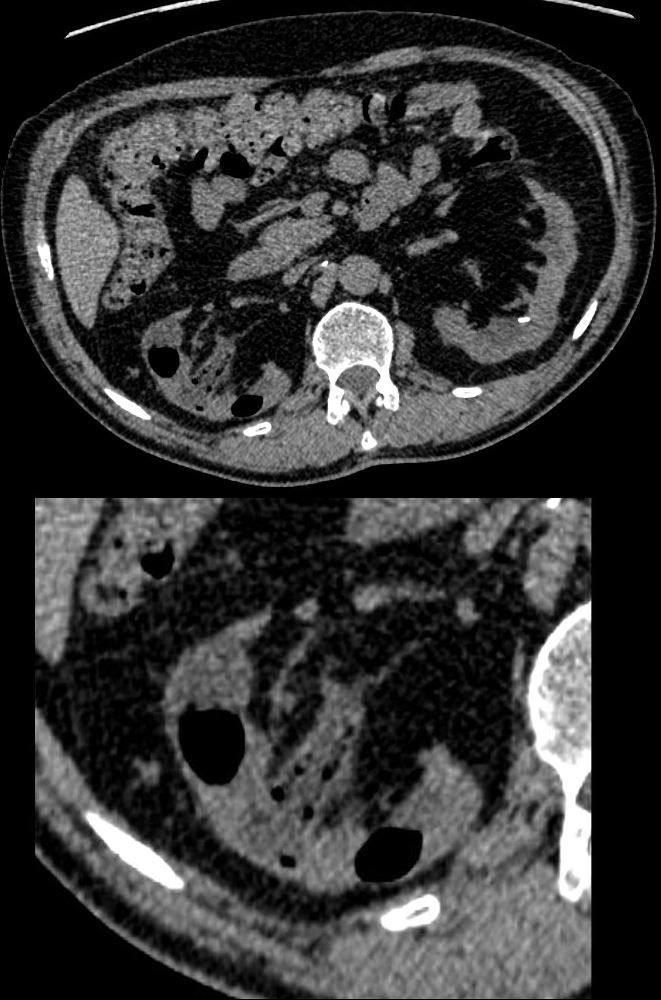
Axial CT Non Contrast– Emphysematous Pyelonephritis
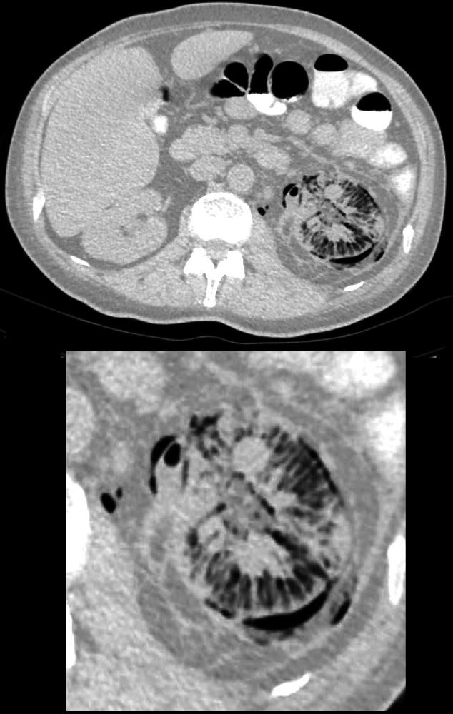
55-year-old diabetic female presents with flank pain and fever. CT shows diffuse infiltration of air into the swollen kidney including the intertstitium of the renal parenchyma, subcapsular regions, perinephric space and the retroperitoneum. .There are islands of soft tissue predominantly in the medial aspect of the kidney. Perinephric induration and thickening of Gerota’s fascia is noted.
Ashley Davidoff MD TheCommonVein.net 135710c
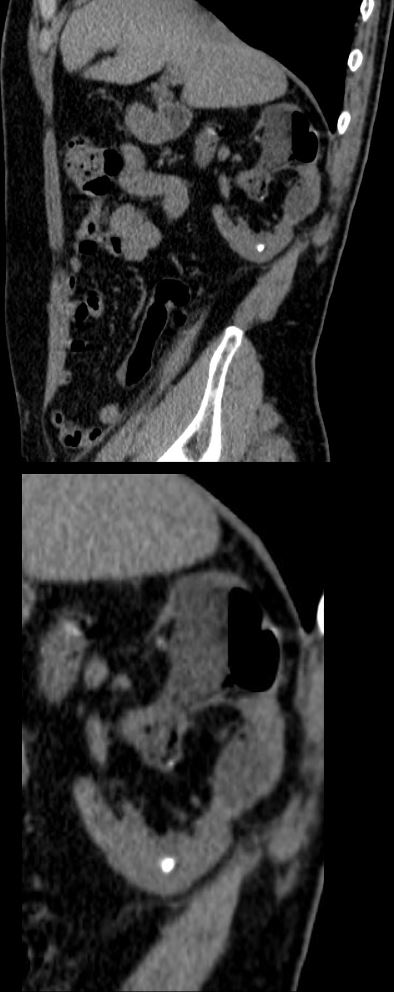
Axial CT with Contrast– Emphysematous Pyelonephritis
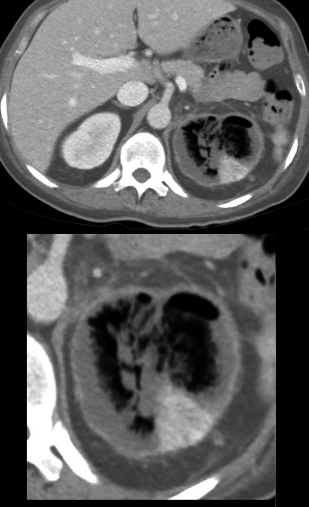
55-year-old diabetic female presents with flank pain and fever. CT with contrast in the nephrogram phase shows a non-functioning rim of cortex and medulla, and a small island of functioning parenchyma in the posterolateral portion of the left kidney. There is a thin enhancing rim that likely represents the enhancing capsule.
Ashley Davidoff MD TheCommonVein.net 135716c
A Second Patient
68-year-old diabetic male in chronic renal failure presents with right flank pain and fever. CT without contrast shows air within the mid and lower portions of the right kidney, with speckled bubbles of air in the parenchyma of the upper portions
Ashley Davidoff MD TheCommonVein.net 135733c
68-year-old diabetic male in chronic renal failure presents with right flank pain and fever. CT without contrast shows air within the parenchyma of the right kidney, with speckled bubbles of air and thick fluid in the renal pelvis
Ashley Davidoff MD TheCommonVein.net 135734c01
68-year-old diabetic male in chronic renal failure presents with right flank pain and fever. CT without contrast shows air within the parenchyma of the right kidney, with speckled bubbles of air and thick fluid in the renal pelvis. Focal calcification also noted in the lower pole. There is overall thinning of the parenchyma.
Ashley Davidoff MD TheCommonVein.net 135735c