
US Sagittal – Posterior Urethral Valves Keyhole Sign
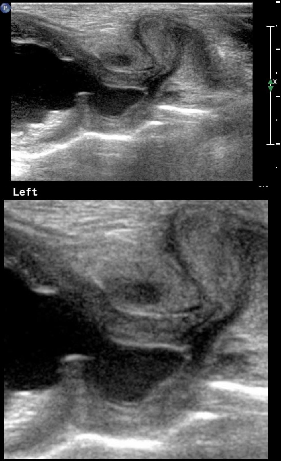
1-day old baby boy who was noted to have hydronephrosis on a prenatal ultrasound. Sagittal view of the thick-walled bladder and dilated proximal urethra is reminiscent of a keyhole. These findings are manifestations of a proximal severe obstruction of the posterior urethra with secondary dilation caused by posterior urethral valves, with secondary bladder outlet obstruction and hypertrophy of the bladder as a result
Ashley Davidoff MD TheCommonVein.net 135747c
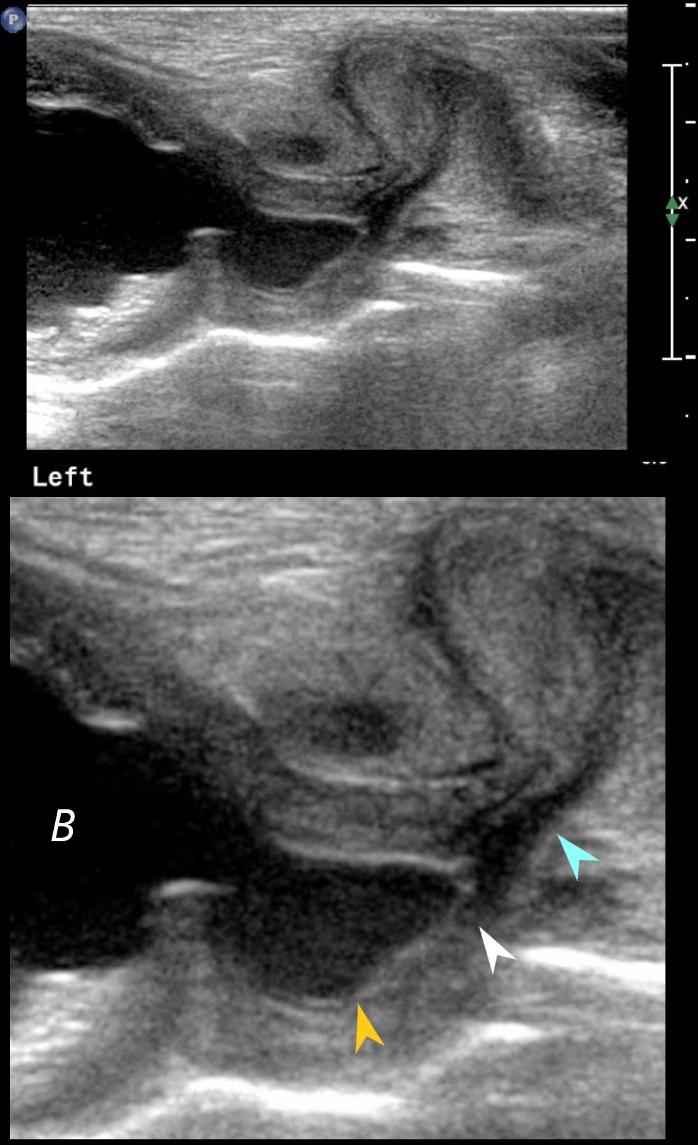
1-day old baby boy who was noted to have hydronephrosis on a prenatal ultrasound. Sagittal view of the thick-walled bladder (B) and dilated proximal urethra (orange arrowhead) is reminiscent of a keyhole. These findings are manifestations of a proximal severe obstruction of the posterior urethra with secondary dilation (orange arrowhead) caused by posterior urethral valves, (white arrowhead), with secondary bladder outlet obstruction and hypertrophy of the bladder as a result . Distal to the obstruction the penile urethra assumes a normal caliber
Ashley Davidoff MD TheCommonVein.net 135747cL
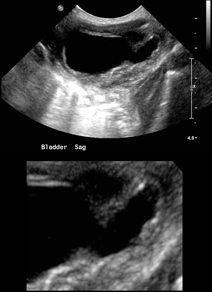
1-day old baby boy who was noted to have hydronephrosis on a prenatal ultrasound. Sagittal view of the thick-walled bladder and dilated proximal urethra is reminiscent of a keyhole. These findings are manifestations of a proximal severe obstruction of the posterior urethra with secondary dilation caused by posterior urethral valves, with secondary bladder outlet obstruction and hypertrophy of the bladder as a result
Ashley Davidoff MD TheCommonVein.net 135743c
Posterior urethral valves (PUV) is a congenital condition that occurs in male infants during fetal development. It is the most common cause of bladder outlet obstruction in newborns. In this condition, there are abnormal valves in the urethra, that can cause obstruction
- Cause: The exact cause of posterior urethral valves is not well understood, but it is thought to result from a developmental abnormality in the fetal urethra during the early stages of pregnancy.
- Incidence: This condition is much more common in males than females, and it is a rare disorder, occurring in about 1 in 5,000 live male births.
- Presentation: PUV is usually diagnosed prenatally or shortly after birth. The typical signs and symptoms include difficulty urinating, a weak urinary stream, urinary tract infections, and an enlarged bladder. hydronephrosis
- Complications: The obstruction can cause the bladder to become thickened and distended, leading to backflow of urine into the ureters and kidneys causing hydroureter and hydronephrosis.
- Diagnosis: Prenatal ultrasound can often detect hydronephrosis, and sometimes detect the presence of posterior urethral valves during pregnancy. After birth, imaging studies such as a voiding cystourethrogram (VCUG) or a renal ultrasound may be used to confirm the diagnosis.
- Treatment: The primary treatment for posterior urethral valves is surgical intervention. The goal of surgery is to remove or ablate the obstructive tissue in the urethra, allowing for proper urine flow. Early diagnosis and treatment are crucial to prevent kidney damage.
- Prognosis: The prognosis for infants with posterior urethral valves depends on the severity of the obstruction and the promptness of intervention. With early diagnosis and appropriate treatment, many children can lead relatively normal lives. However, some may experience long-term issues related to kidney function.
Management often involves a multidisciplinary approach
US Transverse – Thick-Walled Bladder
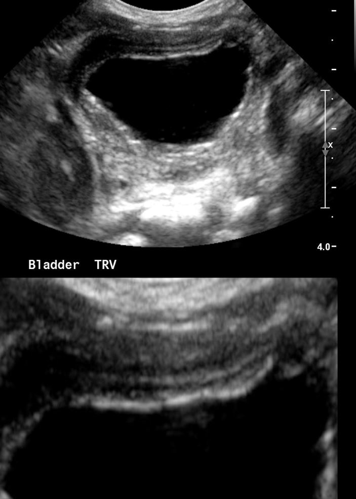
1-day old baby boy who was noted to have hydronephrosis on a prenatal ultrasound. The urinary bladder is noted to be thick-walled, secondary to the bladder outlet obstruction caused by the posterior urethral valves
Ashley Davidoff MD TheCommonVein.net 135751c
US Sagittal – Bilateral Hydronephrosis and Hydroureter
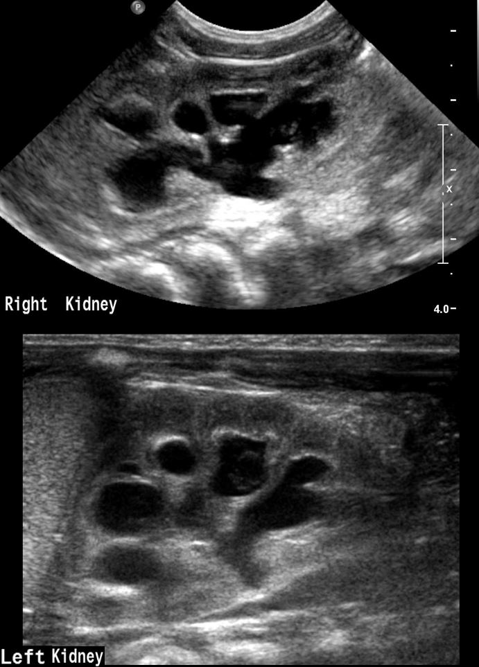
1-day old baby boy who was noted to have hydronephrosis on a prenatal ultrasound. There is bilateral hydronephrosis and hydroureter, secondary to the bladder outlet obstruction caused by the posterior urethral valves
Ashley Davidoff MD TheCommonVein.net 135752c
5 year Old – Voiding Cystourethrogram –
Posterior Urethral Valves – Keyhole Sign
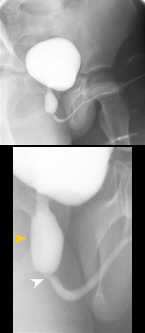
5-year-old boy who was noted to have hydronephrosis on ultrasound. VCUG in the RAO oblique-lateral view shows dilated proximal urethra (orange arrowhead) caused by posterior urethral valves, (white arrowhead), with secondary bladder outlet obstruction. The resulting shapes are reminiscent of a keyhole – and hence the “keyhole sign”. Distal to the obstruction the penile urethra assumes a normal caliber
Ashley Davidoff MD TheCommonVein.net 135754cL
Voiding Cystourethrogram –
Posterior Urethral Valves – Bladder Diverticula
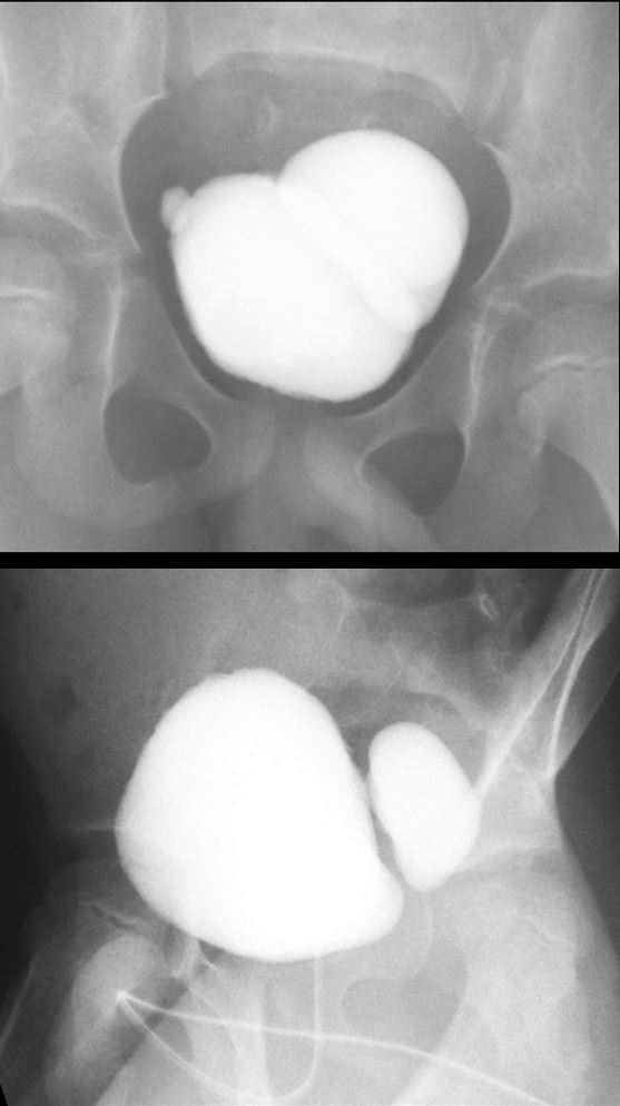
5-year-old boy who was noted to have hydronephrosis on ultrasound. VCUG shows a contrast filled bladder with secondary bladder outlet obstruction and bilateral diverticula. The right diverticulum is small while the left is very large.
Ashley Davidoff MD TheCommonVein.net 135756c
US Transverse and Sagittal – Bladder Diverticulum
Secondary to Bladder Outlet Obstruction
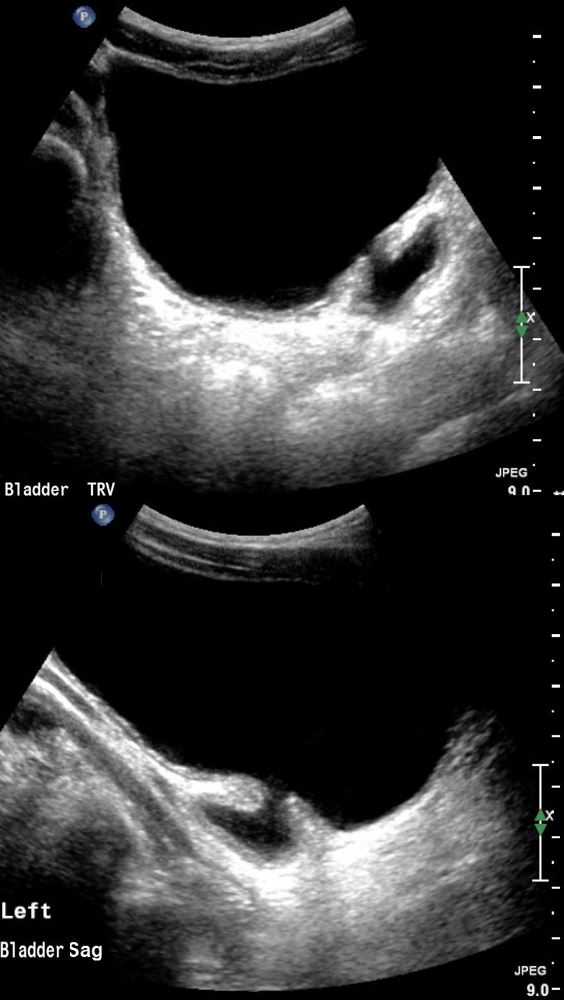
5-year-old boy who was noted to have hydronephrosis on ultrasound. US in transverse and sagittal projections shows a left sided bladder diverticulum secondary to bladder outlet obstruction from posterior urethral valves.
Ashley Davidoff MD TheCommonVein.net 135757c