
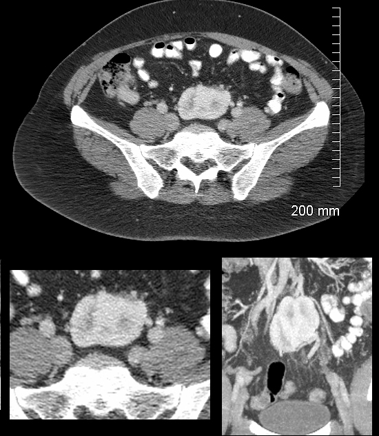
CT in the axial plane shows a reniform structure in the presacral region between the cortico-medullary and nephrographic phase of the contrast enhanced study. The bottom left image is a magnification view of the axial CT. The bottom right image is in the coronal plane and shows a maloriented pelvic kidney in the same recognizable phase of renal contrast excretion. A normal kidney was noted in the right renal fossa and the diagnosis is consistent with a pelvic kidney
Ashley Davidoff MD TheCommonVein.net 132055c