
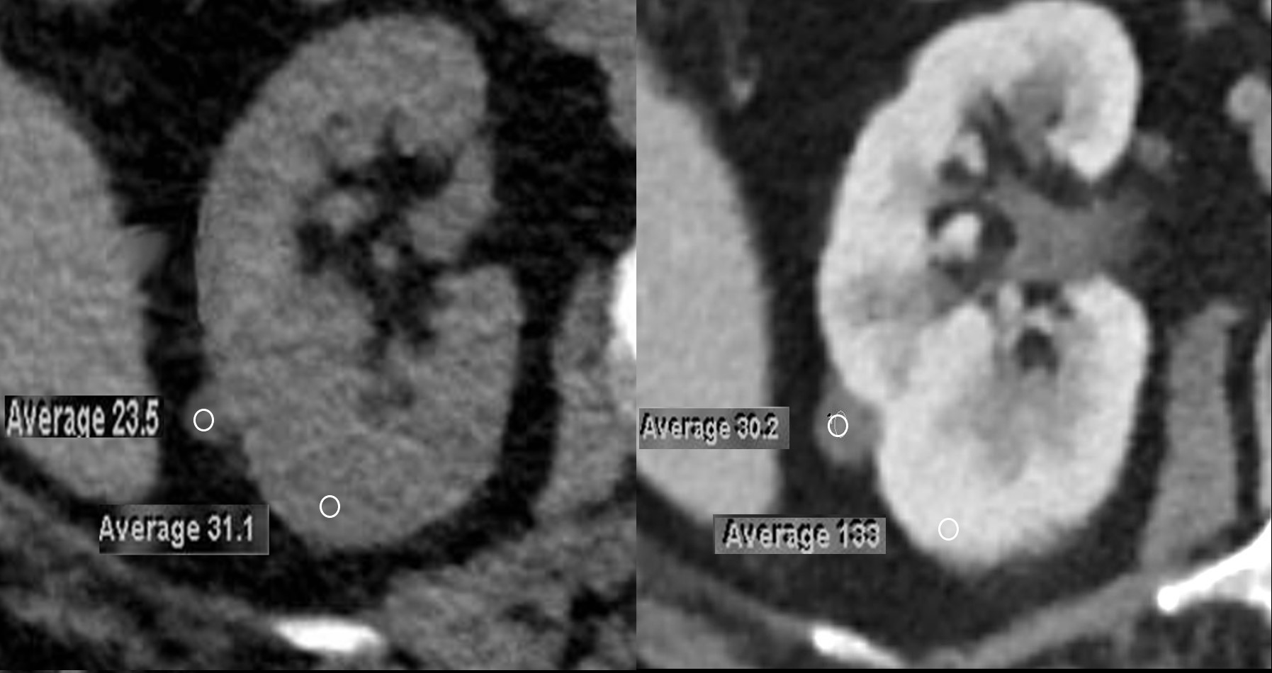
72-year-old female presents with abdominal pain. A 9mm homogeneously hyperdense cystic appearing lesion is noted on the non-contrast CT and it measures 23.5HU and is consistent with an hemorrhagic cyst or proteinaceous cyst. The lesion measures30HU following contrast (SD 10HU) in the late corticomedullary phase. The normal kidney measures 31.1HU before contrast and 133HU after contrast
Ashley Davidoff MD TheCommonVein.net 136019cL
Coronal CT Hemorrhagic Cyst of the Kidney Post Contrast
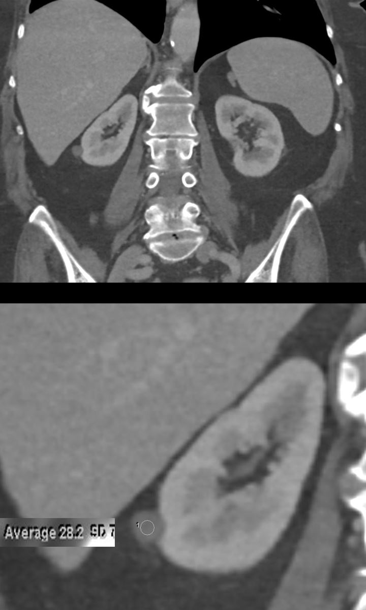
72-year-old female presents with abdominal pain. On a non-contrast CT, the 10mm homogeneously hyperdense cystic lesion measured 23.5HU. The lesion measures 28.2HU following contrast (SD 10HU) in the late corticomedullary phase. The normal kidney measures 31.1HU before contrast and 133HU after contrast
Ashley Davidoff MD TheCommonVein.net 136027c
High Density Hemorrhagic or Proteinaceous Cyst Non Contrast CT
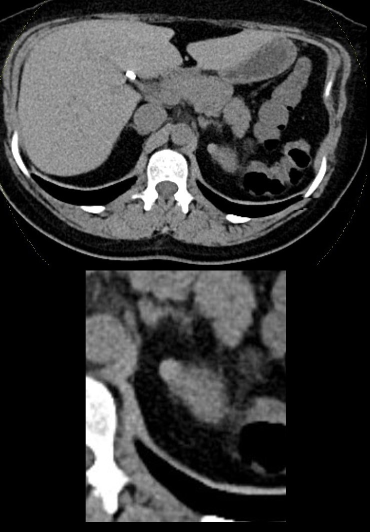
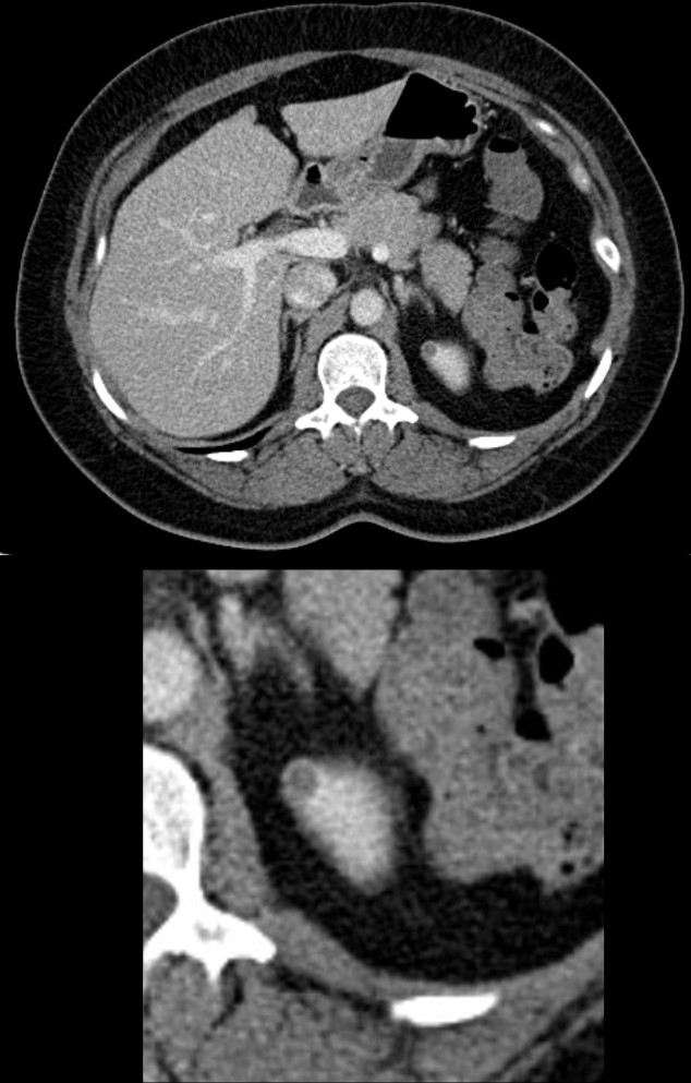
Non-Contrast CT in the axial plane shows an 8mm homogeneous hyperdense lesion, exophytic off the upper pole of the left kidney. A hyperdense cyst as a result of hemorrhage or accumulation of proteinaceous material are most likely
Ashley Davidoff MD TheCommonVein.net TCV 24K 135914c
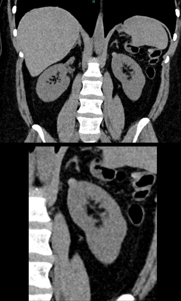
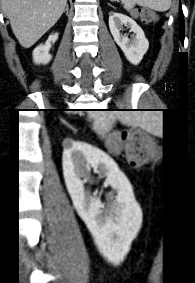
Non-Contrast CT in the coronal plane shows an 8mm homogeneous hyperdense lesion, exophytic off the upper pole of the left kidney. A hyperdense cyst as a result of hemorrhage or accumulation of proteinaceous material are most likely. The right kidney has a deformity of the upper pole secondary to atrophy of the upper pole moiety of a duplicated collecting system
Ashley Davidoff MD TheCommonVein.net TCV 24K 135915c
CT with Contrast
70 Second Delay in Late Corticomedullary Phase
CT in the axial plane shows an 8mm homogeneous that has now become relatively hypodense, exophytic off the upper pole of the left kidney. It does not appear to enhance, though it does appear to have a hyper-enhancing wall, which more likely relates to normal surrounding parenchyma. .
A hemorrhagic or proteinaceous cyst is most likely. MRI subsequently confirmed the diagnosis
Ashley Davidoff MD TheCommonVein.net TCV 24K 135918c
CT in the coronal plane shows an 8mm homogeneous that has now become relatively hypodense, exophytic off the upper pole of the left kidney. It does not appear to enhance.
A hemorrhagic or proteinaceous cyst is most likely. MRI subsequently confirmed the diagnosis
Ashley Davidoff MD TheCommonVein.net TCV 24K 135919c
CT with Contrast 6minute Delay in Pyelographic(Excretory) Phase
High Density Hemorrhagic or Proteinaceous Cyst of the Upper Pole of the Left Kidney
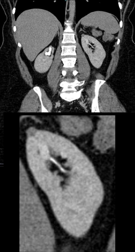
CT in the coronal plane shows an 8mm homogeneous that remains relatively hypodense, exophytic off the upper pole of the left kidney. .A hemorrhagic or proteinaceous cyst is most likely. MRI subsequently confirmed the diagnosis
The right kidney shows contrast in a mildly dilated collecting system in an upper pole moiety with secondary atrophy of the upper pole moiety of a duplicated collecting system
Ashley Davidoff MD TheCommonVein.net TCV 24K 135920c
Pre Contrast T1 Weighted MRI –
T1 Bright Hemorrhagic or Proteinaceous Cyst
T1 weighted MRI in the axial plane shows a T1 bright 8mm homogeneous lesion, exophytic off the upper pole of the left kidney.
This finding is compatible with a hemorrhagic or proteinaceous cyst
Ashley Davidoff MD TheCommonVein.net TCV 24K 135921c
In and Out of Phase Pre-Contrast T1-Weighted MR
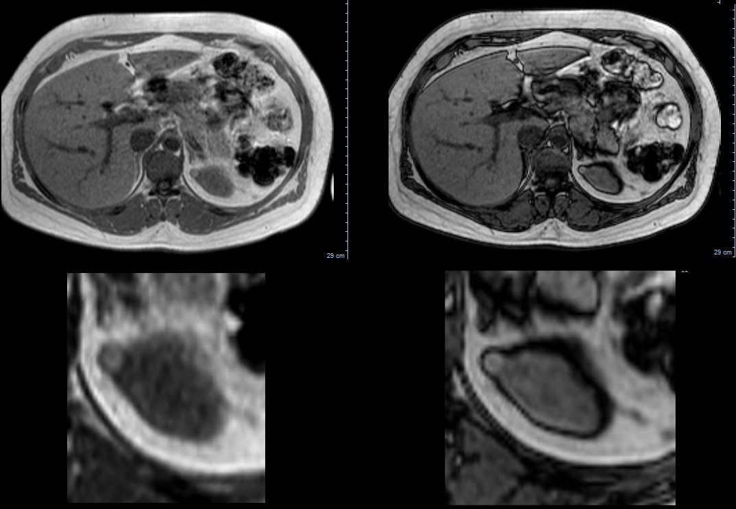
T1 weighted MRI in the axial plane shows a T1 bright 8mm homogeneous lesion, exophytic off the upper pole of the left kidney.
This finding is compatible with a hemorrhagic or proteinaceous cyst
Ashley Davidoff MD TheCommonVein.net TCV 24K 135923c
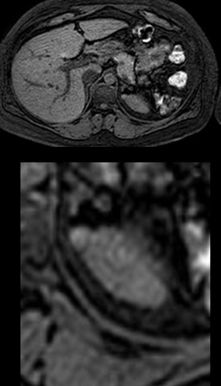
Contrast Enhanced T1 Weighted Image in the Arterial Phase-
No Internal Enhancement
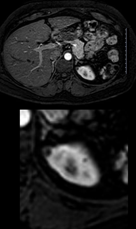
Contrast enhance MRI in the axial plane at 20seconds shows no enhancement of the matrix of the cyst. The wall does enhance but is thin.
This finding is compatible with a hemorrhagic or proteinaceous cyst
Ashley Davidoff MD TheCommonVein.net TCV 24K 135926c
Subtraction of a Contrast Enhanced T1 Weighted Image in the Arterial Phase-No Internal Enhancement
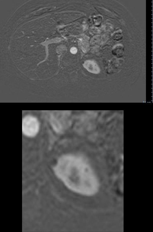
Subtraction technique in the axial plane at 20seconds shows no enhancement of the matrix of the cyst. The wall does enhance minimally but is thin and smooth.
This finding is compatible with a hemorrhagic or proteinaceous cyst
Ashley Davidoff MD TheCommonVein.net TCV 24K 135927c