Nephrolithiasis Pre-lithotripsy
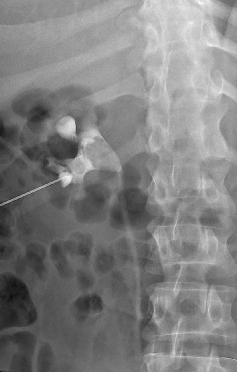
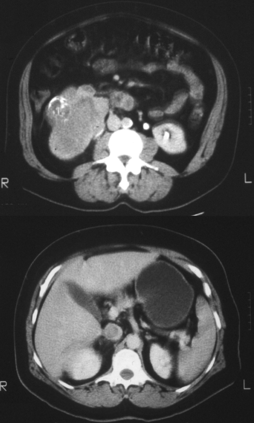
37year-old male presents with back pain Antegrade pyelogram shows 2 filling defects in the renal pelvis with the larger occupying almost the entire downstream pelvis. There is mild hydronephrosis. Contrast is seen in the ureter
Ashley Davidoff MD TheCommonVein.net 135460
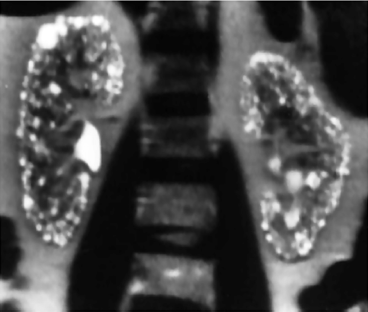
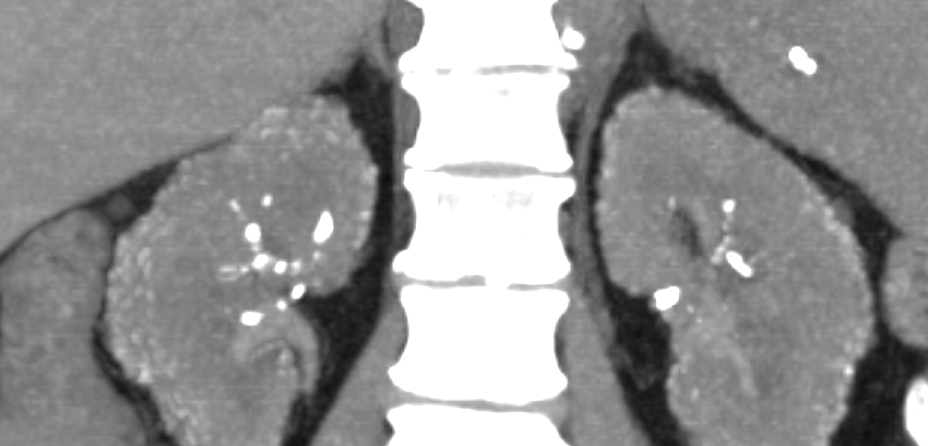
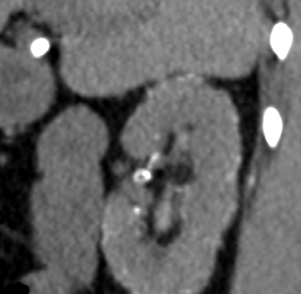
37year-old male presents with back pain Non contrast CT shows 2 calcifications. The larger stone is in the renal pelvis and the smaller in a lower pole calyx.
Ashley Davidoff MD TheCommonVein.net 135461
Cancer
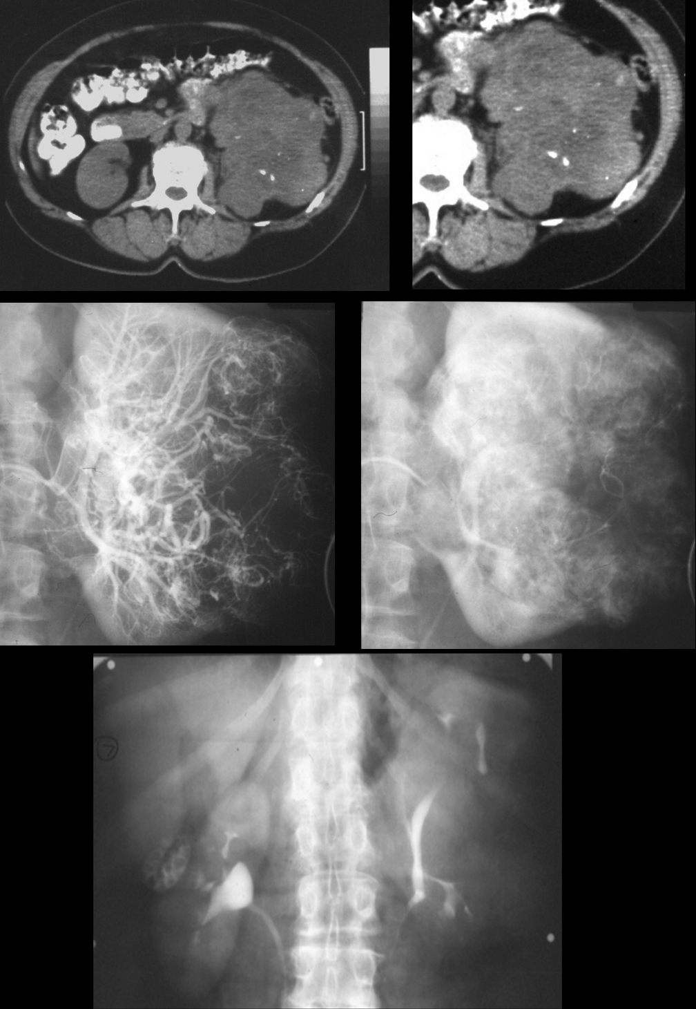
On CT there is dystrophic calcification. On angiography there is hypervascularity, neovascularity, AV shunting. Excretory phase after angiography shows distorted calyces
Ashley Davidoff MD
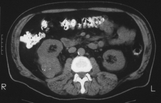
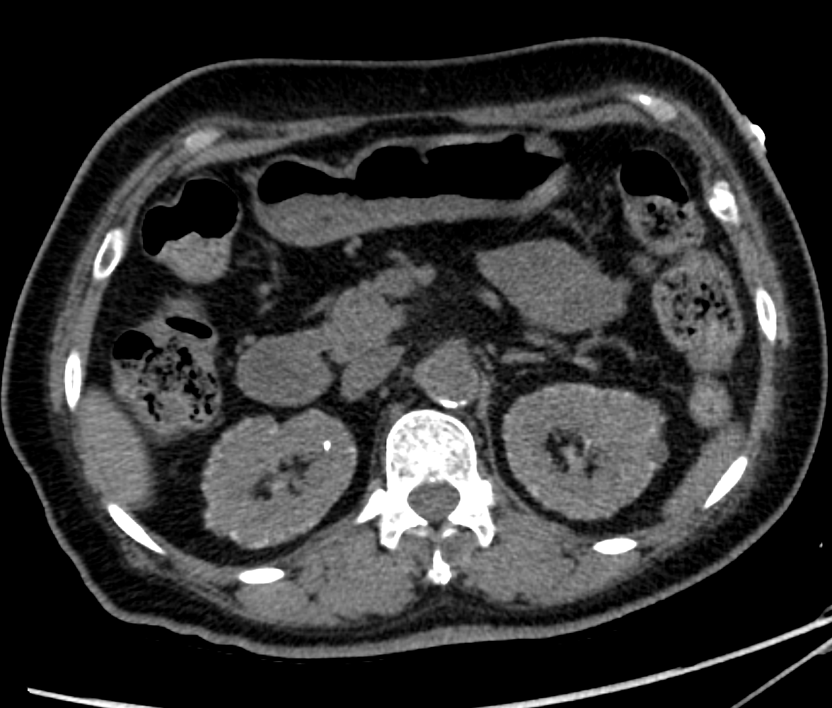
Polycystic Kidney Disease PCKD
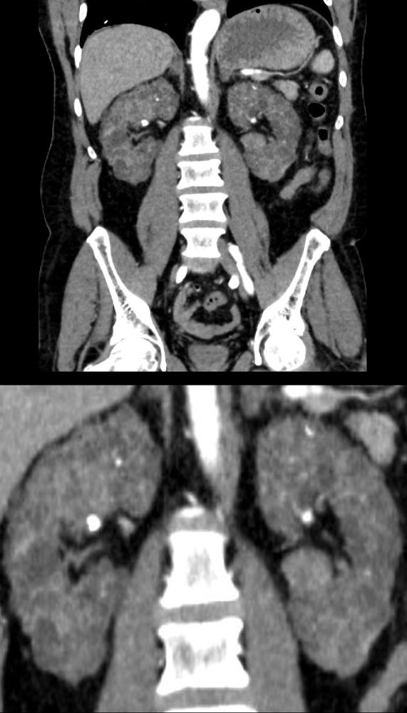
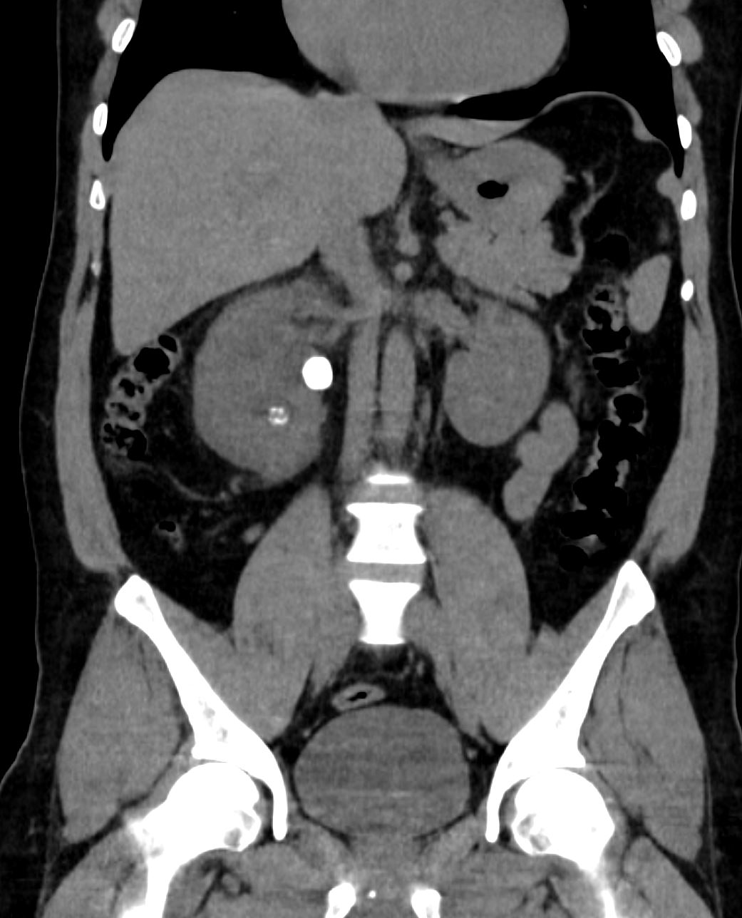
56-year-old male in chronic renal failure presents with flank pain. CT with contrast shows diffuse cystic changes of near normal sized kidneys with total replacement of the renal parenchyma by small uniformly sized cysts. Multiple calcifications are scattered throughout the parenchyma.
Ashley Davidoff MD TheCommonVein.net 135723c
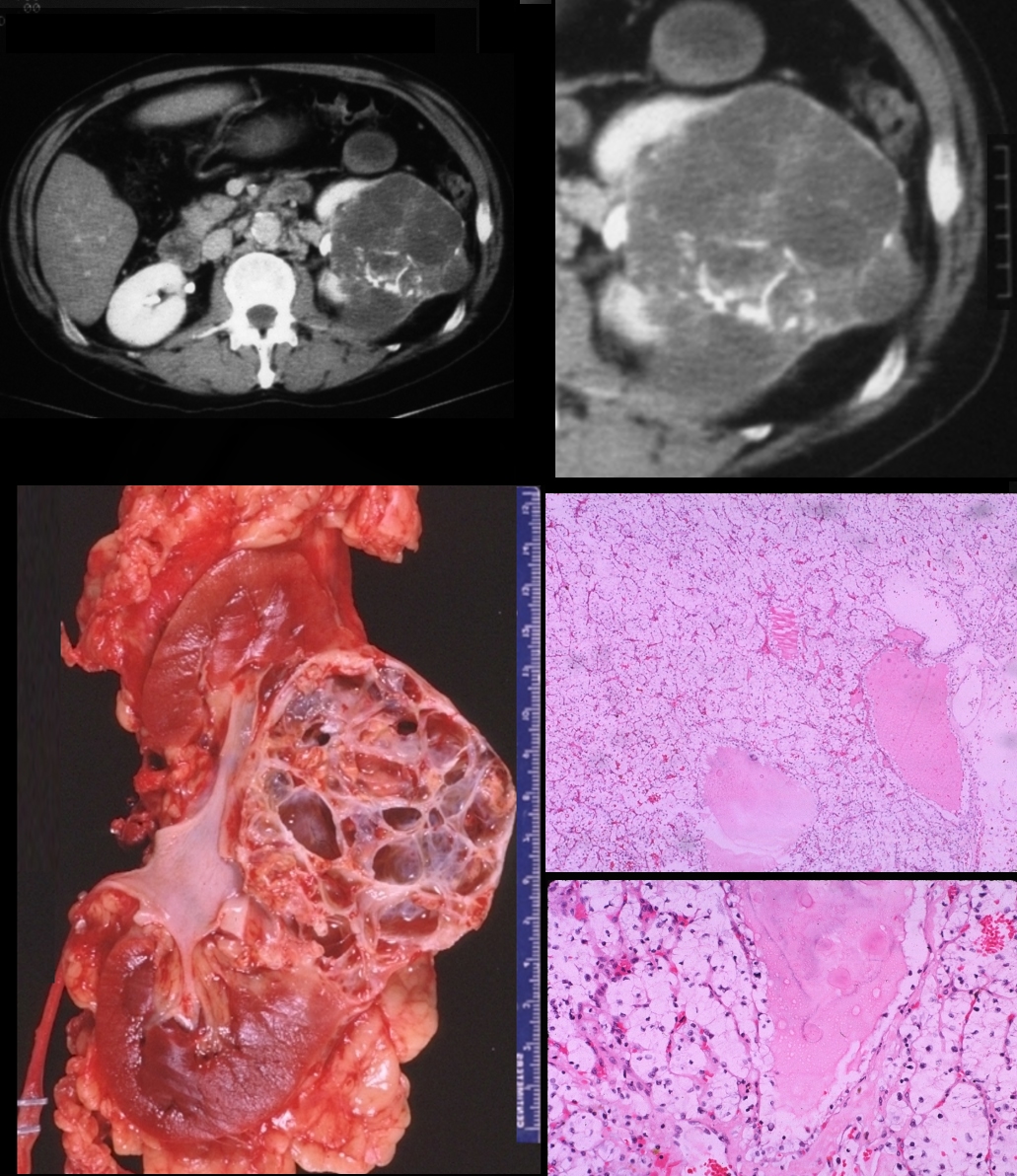
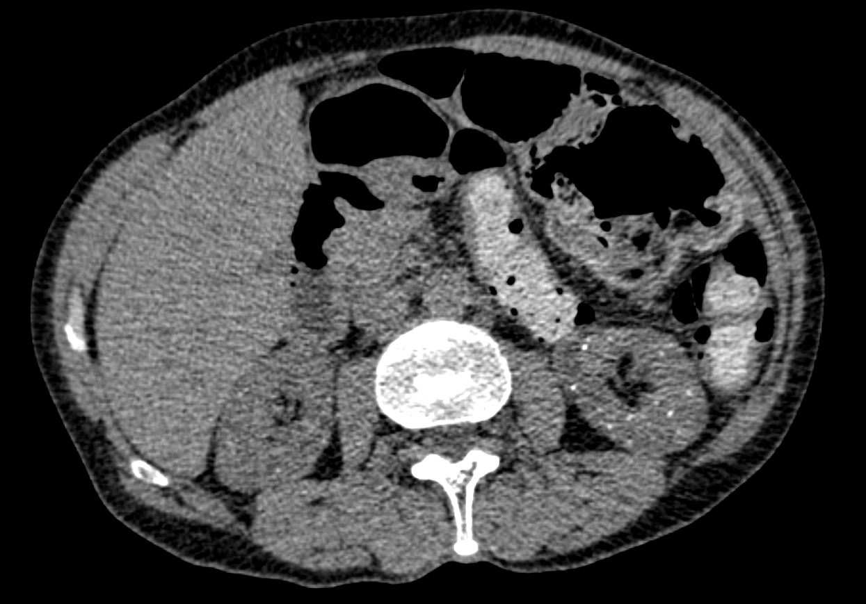
Cystic renal cell carcinoma with dystrophic calcification in the mid portion of the left kidney. Pathology reveals cystic renal cell carcinoma with clear cell histopathology
Ashley Davidoff MD
Ashley Davidoff MD
Ashley Davidoff MD
Ashley Davidoff MD
Ashley Davidoff MD
Ashley Davidoff MD
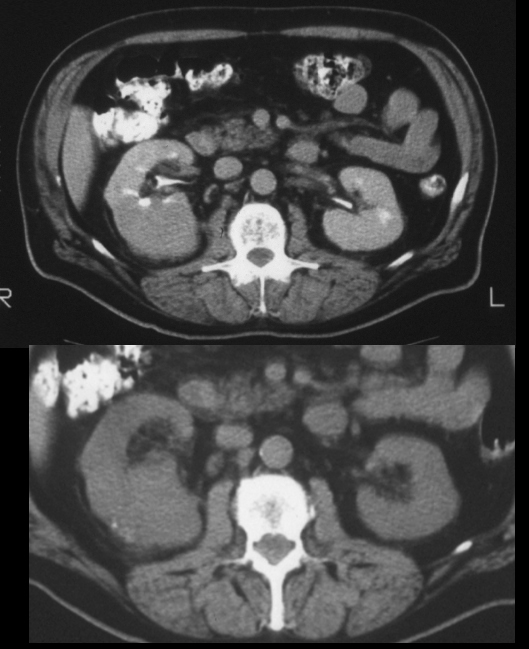
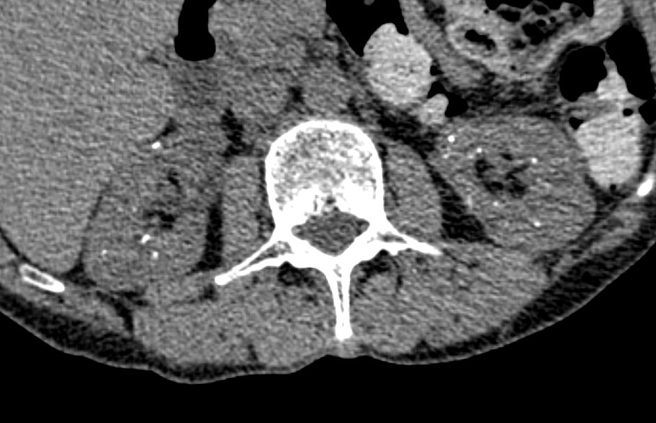
Metabolic Causes
Lithium
CT shows numerous punctate calcifications in the cortex
and medulla bilaterally. Given history of lithium usage, CT appearance can be
seen with lithium-induced renal disease.
Ashley Davidoff The CommonVein.net
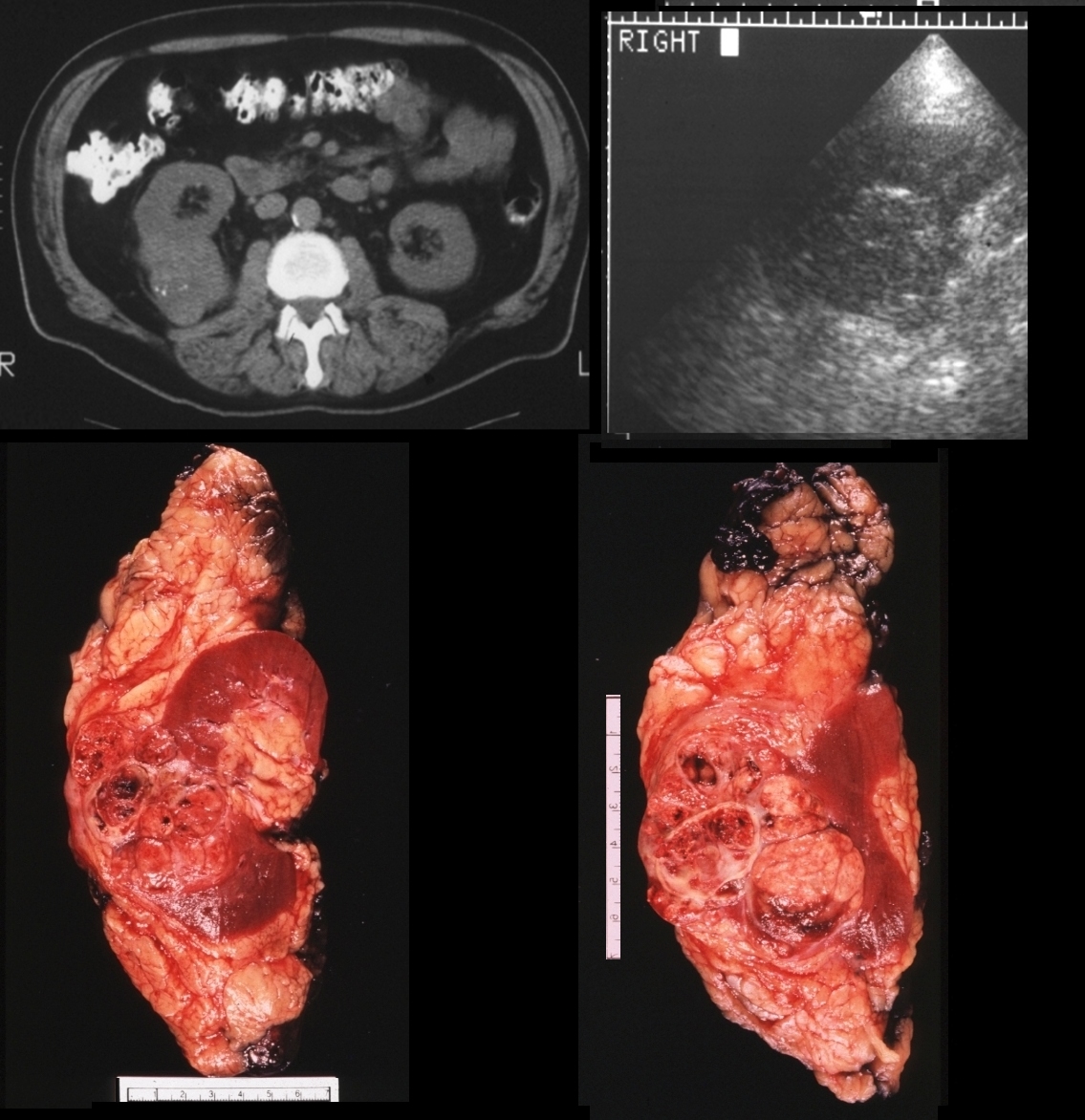
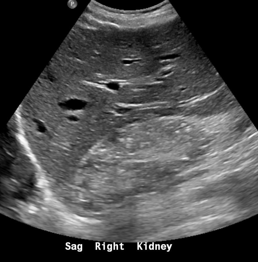
CT shows numerous punctate calcifications in the cortex
and medulla bilaterally. Given history of lithium usage, CT appearance can be
seen with lithium-induced renal disease.
Ashley Davidoff The CommonVein.net
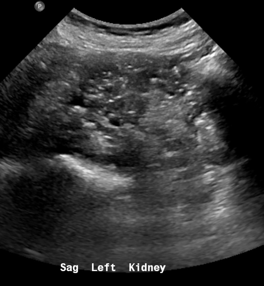
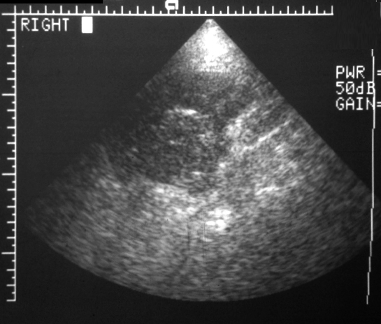
and medulla. CT showed multiple punctate calcifications Given history of lithium usage, sonographic appearance can be
seen with lithium-induced renal disease.
Ashley Davidoff The CommonVein.net
Consequence of lithium therapy is the most likely diagnosis
Ashley Davidoff MD
018 72F Asian renal capsular calcification
Consequence of lithium therapy is the most likely diagnosis
Ashley Davidoff MD
019 72F Asian renal capsular calcification
Consequence of lithium therapy is the most likely diagnosis
Ashley Davidoff MD
020 72F Asian renal capsular calcification