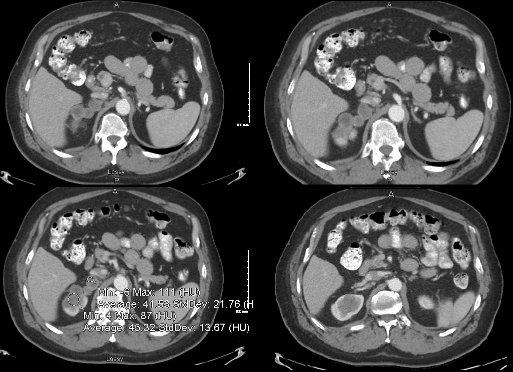
58 year old male with a relatively low density (41HU) lobulated mass originating from the upper pole of the right kidney. Pathology revealed a diagnosis of a benign Schwannoma
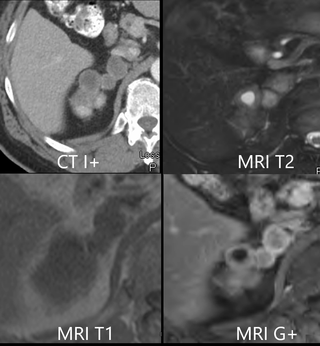
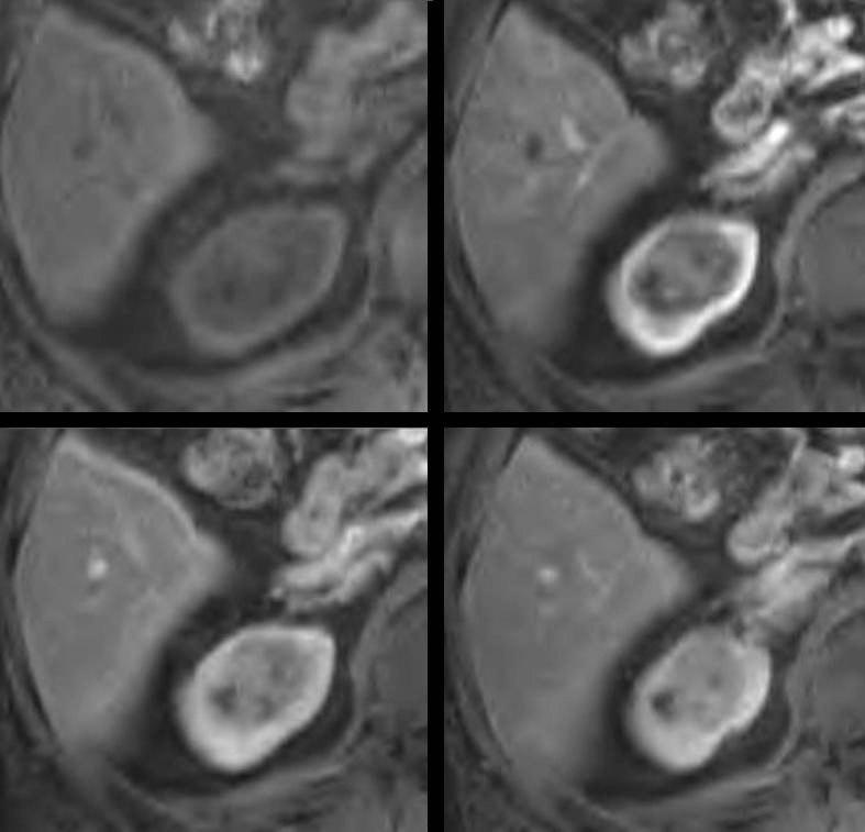
Axial Imaging CT and MRI
Collage of a axial imaging through a right upper pole mass in a 58 year old male showing a lobulated mass, varicoid in shape. using CT and MRI The left upper image shows a contrast enhanced CT with relatively low density matrix (41HU) and enhancing thick smooth wall. A T2 weighted image (left upper) shows a combination of T2 brightness and intermediate signal. The left lower image is a T1 weighted image showing iso-intensity of the mass with other soft tissues while the contrast enhanced MRI shows enhancing walls (41HU) lobulated mass originating from the upper pole of the right and a combination in the matrix of the lobules of dark signal and intermediate signal. Pathology revealed a diagnosis of a benign Schwannoma Ashley Davidoff MD TheCommonVein.net
CT scan in the Axial Plane
Axial Imaging MRI T2 Weighted
T2 weighted MRI of a 58 year old male lobulated mass originating from the upper pole of the right kidney showing components of T2 brightening . The mass shows a combination of T2 brightness . The nodule closest to the superior pole of the kidney (white arrowhead) has a very bright central component (best seen in the right lower image) . It is as bright as the CSF and likely is cystic in nature It is associated with a thick wall. The other clustered nodular components abutting the vertebral body (blue arrowheads) are less bright and less well defined. Pathology revealed a diagnosis of a benign Schwannoma Ashley Davidoff TheCommonVein.net
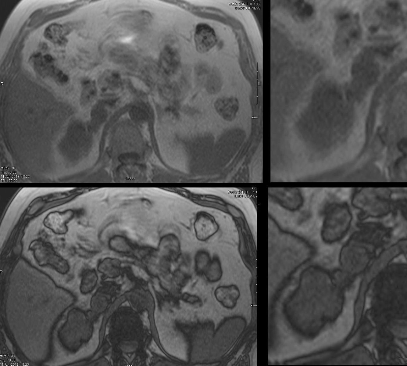
Axial Imaging MRI T1 Weighted
MRI imaging with in (upper images) and out of phase T1 weighted imaging (lower images) shows no darkening in the out of phase images. Pathology revealed a diagnosis of a benign Schwannoma Ashley Davidoff MD TheCommonVein.net
MRI in the Axial Plane Gadolinium Enhanced Weighted Images
Gadolinium enhanced MRI in the arterial phase in a 58 year old male confirms a lobulated varicoid mass originating from the upper pole of the right kidney . The nodule closest to the superior pole of the kidney (white arrowhead) is dark and does not enhance and is likely a cysts, with thickened wall. The other clustered nodular components abutting the vertebral body (blue arrowheads) have bright enhancement of the wall and mild diffuse enhancement of the matrix. Pathology revealed a diagnosis of a benign Schwannoma Ashley Davidoff TheCommonVein.net
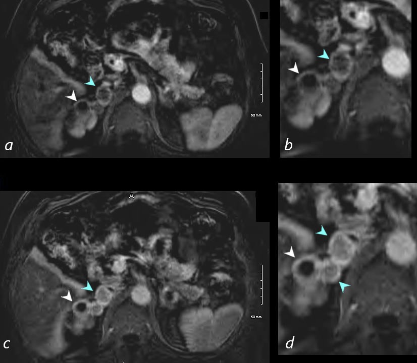
Pre-Contrast Arterial Phase and Delayed in Axial Plane
The MRI in the axial plane shows the base of the mass at its origin from the right kidney prior to (a) during the arterial phase of contrast administration (b) and on delay (c and d) Prior to the Gadolinium injection the mass on T1 weighted imaging is relatively dark (white arrowhead, a) compared to the brighter ring of kidney noted (red arrowhead, a). In the arterial phase the kidney enhances (red arrowhead, b) and there is some mild enhancement of the base of the mass (white arrowhead, b) Mild progressive enhancement of the mass continues on the delayed imaging (white arrowheads c and d) . Pathology revealed a diagnosis of a benign Schwannoma Ashley Davidoff TheCommonVein.net
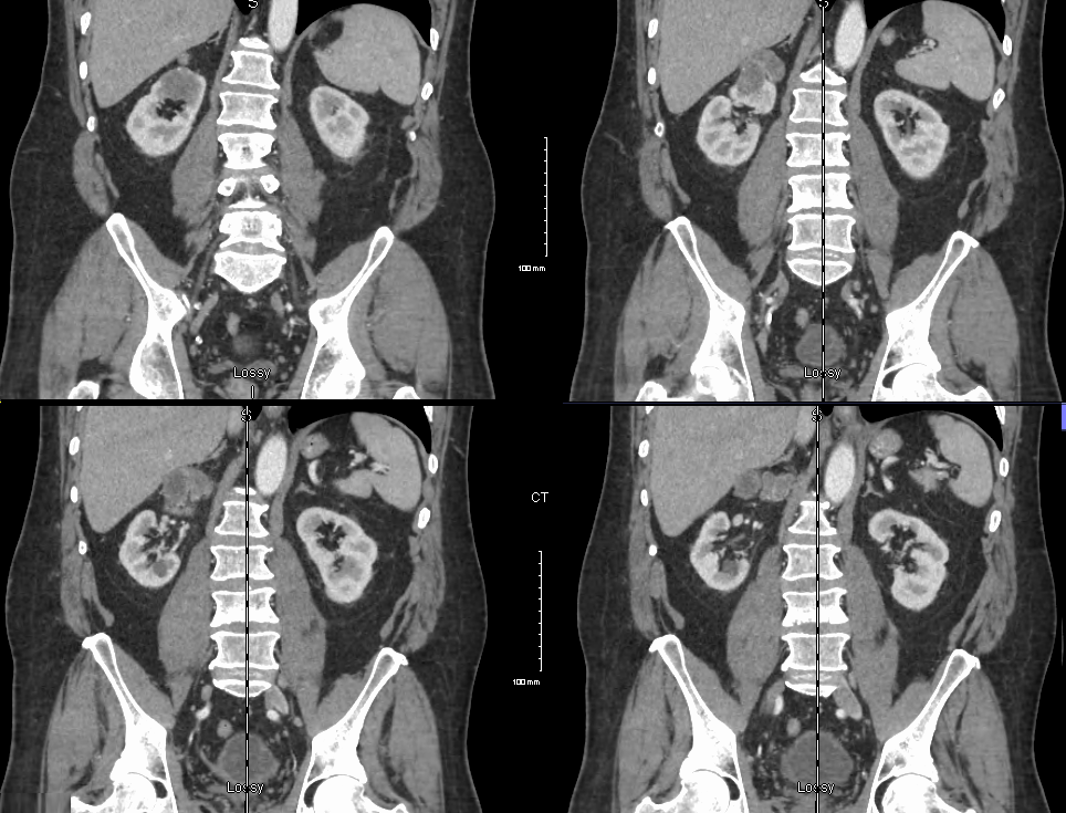
Coronal Imaging CT and MRI
Coronal imaging using contrast enhanced CT (above), and T2 weighted MRI (below) in a 58 year old male shows a relatively low density (41HU) lobulated mass originating from the upper pole of the right kidney, with mild enhancing rim. The T2 weighted image below shows a high intensity T2 weighted component and a superior component with a more heterogeneous appearance with lesser hyperintensity.. Pathology revealed a diagnosis of a benign Schwannoma Ashley Davidoff MD TheCommonVein.netCT scan following contrast of a 58 year old male with a relatively low density (41HU) lobulated mass originating from the upper pole of the right kidney. Pathology revealed a diagnosis of a benign Schwannoma Ashley Davidoff MD TheCommonVein.netT2 weighted coronal MRI of a 58 year old male lobulated mass originating from the upper pole of the right kidney showing components of T2 brightening . Pathology revealed a diagnosis of a benign Schwannoma Ashley Davidoff MD TheCommonVein.net
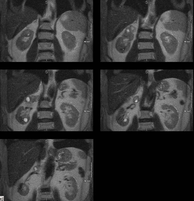
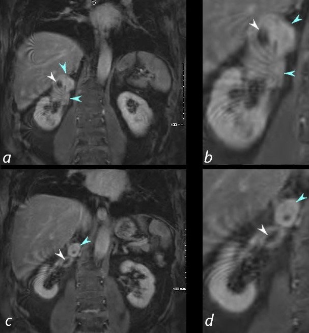
Post Gadolinium in Equilibrium Phase Diffuse Enhancement
Post Gadolinium in Equilibrium Phase Diffuse Enhancement The MRI in the coronal plane shows the mass at in the upper pole of the right kidney delayed enhancement a and b, magnified in c and d) (c and d) There is mild diffuse enhancement of the mass (blue arrowheads, with non enhancement of the cystic component (white arrowhead) Pathology revealed a diagnosis of a benign Schwannoma Ashley Davidoff TheCommonVein.net
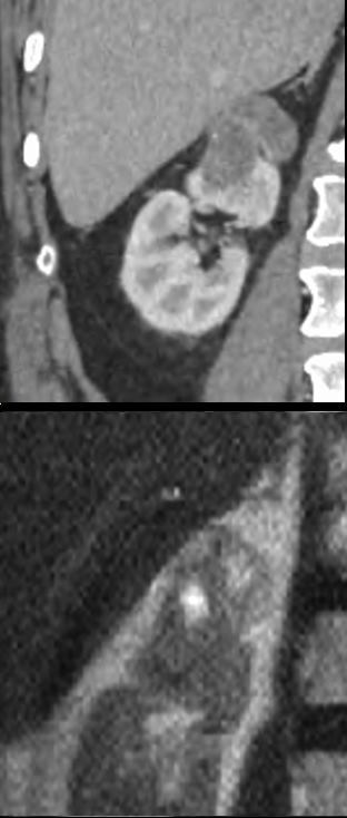
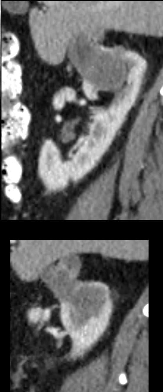
Sagittal CT
CT scan following contrast of a 58 year old male with a relatively low density (41HU) lobulated mass originating from the upper pole of the right kidney. Pathology revealed a diagnosis of a benign Schwannoma Ashley Davidoff MD TheCommonVein.net
Schwannomas are typically benign nerve sheath tumors that arise from the Schwann cells, which produce the myelin sheath that covers nerves. These tumors can develop in any nerve throughout the body.
Regarding the MRI characteristics of Schwannomas, they are generally known to appear as well-defined masses with low to intermediate signal intensity on T1-weighted images and high signal intensity on T2-weighted images. They typically enhance with contrast. However, they are not expected to contain fat.