
Renal Failure Bilateral
Renal Artery Stenosis CT Striated Nephrogram
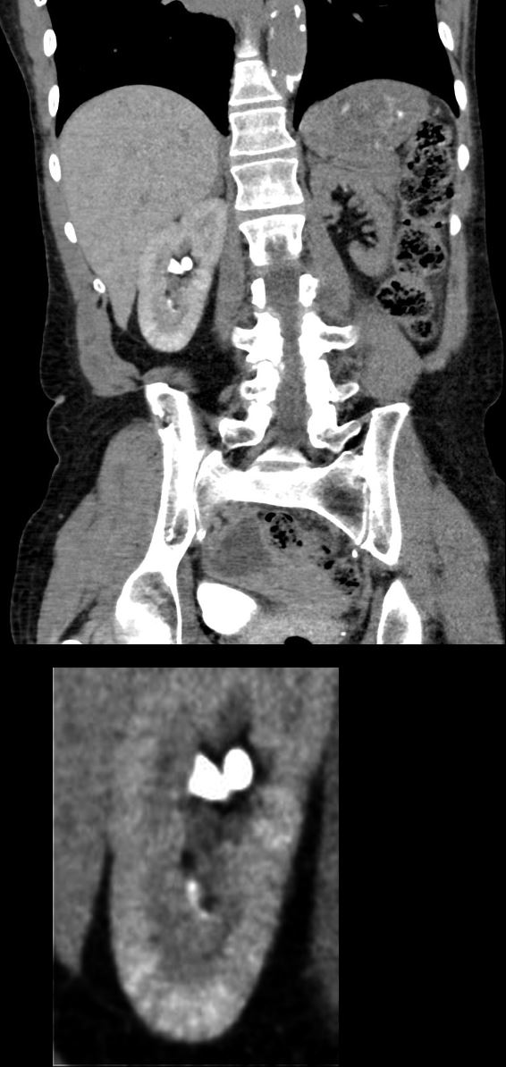
CT scan in the coronal plane in a 61year old female with bilateral severe renal artery calcific atherosclerosis shows a right kidney that is in excretory phase (contrast noted in the upper pole) with an upper pole parenchyma that is in nephrographic phase and a lower pole demonstrates a striated nephrogram (magnified in the lower panel). The left kidney is non-functioning. Part of a contrast filled bladder is noted in the right lower quadrant
Ashley Davidoff MD TheCommonVein.net 013Lu 136068c
CT – Striated Nephrogram Obstructed Kidney Hydronephrosis
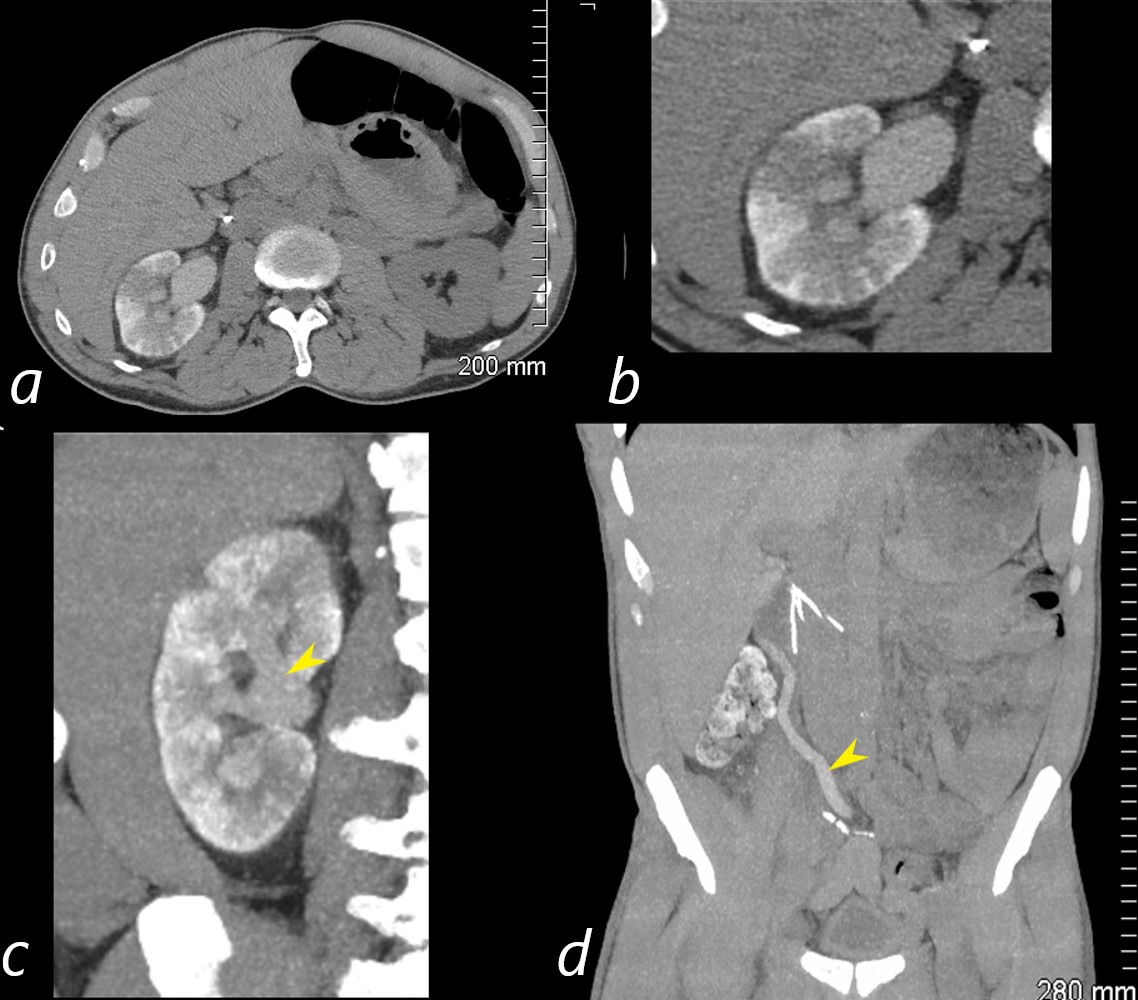
CT scan in the axial plane on delayed imaging a few hours following contrast administration delay shows a persistent striated nephrogram in the right kidney (a,b) ) while the left normal kidney is devoid of contrast after the long delay. The lower panel in the coronal plane (c) shows moderate hydronephrosis, again demonstrates the striated nephrogram but also shows dilute contrast in the dilated collecting system. The right ureter is dilated (d yellow arrowhead) and appears to obstruct at a level of anastomosis (surgical clips)
Ashley Davidoff MD TheCommonVein.net 013Lu 136068c
L