
- 55-year-old female
- past medical history of Type 2 DM
- (with refusal of medications)
- past medical history of Type 2 DM
- `three days of RUQ and LLQ abdominal pain
- associated with
- overall weight loss,
- nausea, and vomiting for the past .
- Labs
- leukocytosis,
- hyperglycemia of 523, and
- Na level of 118 (128 corrected).
Coronal Non Contrast CT– Emphysematous Pyelonephritis
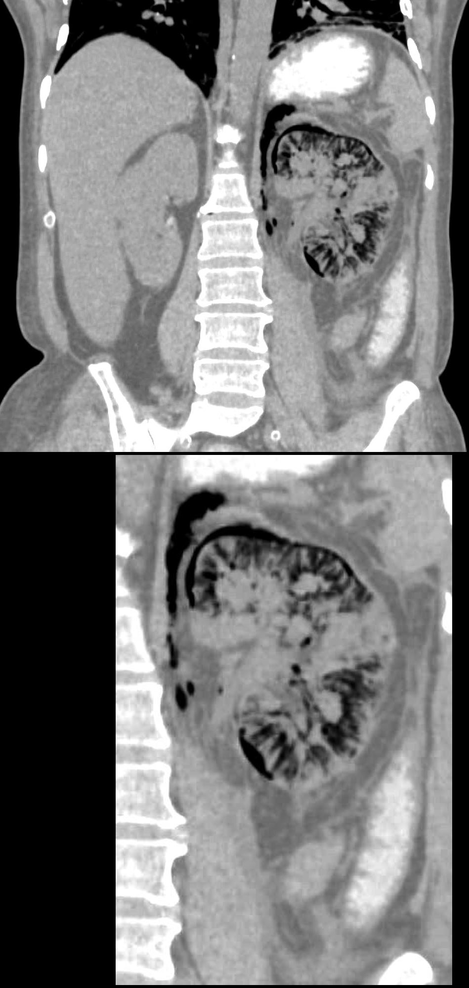
55-year-old diabetic female presents with flank pain and fever. CT without shows diffuse infiltration of air into the swollen left kidney including the intertstitium of the renal parenchyma, subcapsular regions, perinephric space and the retroperitoneum. There is perinephric induration and thickening of Gerota’s fascia and perinephric edema. Small islands of tissue are noted in the upper medial portions of the kidney and the mid portion
Ashley Davidoff MD TheCommonVein.net 135714
Coronal CT with Contrast – Island of Functioning Tissue
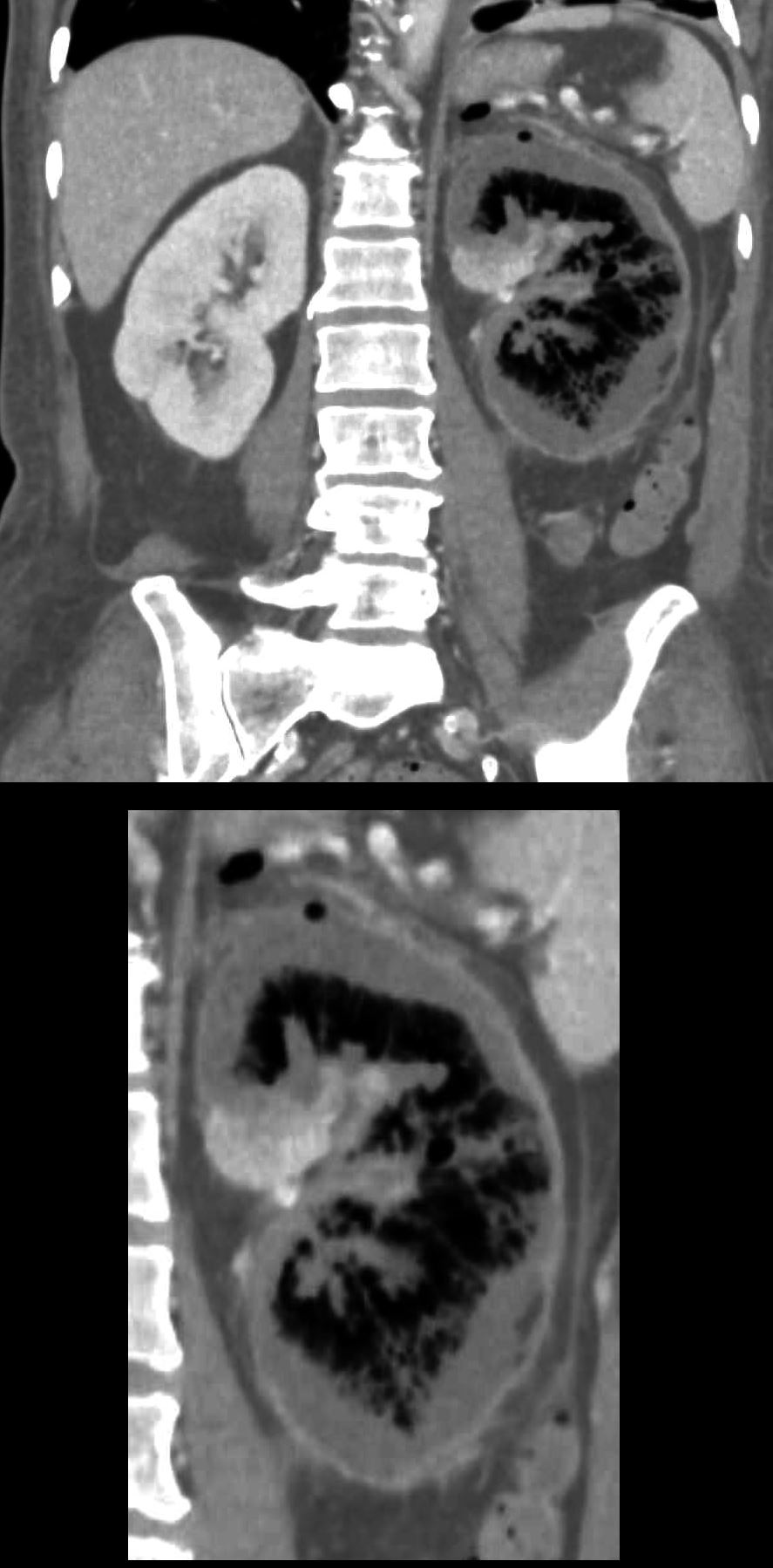
55-year-old diabetic female presents with flank pain and fever. CT with contrast in the nephrogram phase shows a non-functioning rim of cortex and medulla, and a small island of functioning parenchyma in the medial upper portion of the left kidney. There is a thin enhancing rim likely representing an enhancing capsule.
Ashley Davidoff MD TheCommonVein.net 135715c
Axial Non Contrast CT– Emphysematous Pyelonephritis
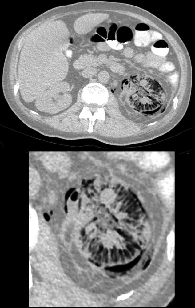
55-year-old diabetic female presents with flank pain and fever. CT shows diffuse infiltration of air into the swollen kidney including the intertstitium of the renal parenchyma, subcapsular regions, perinephric space and the retroperitoneum. .There are islands of soft tissue predominantly in the medial aspect of the kidney. Perinephric induration and thickening of Gerota’s fascia is noted.
Ashley Davidoff MD TheCommonVein.net 135710c
Axial CT with Contrast – Island of Functioning Tissue
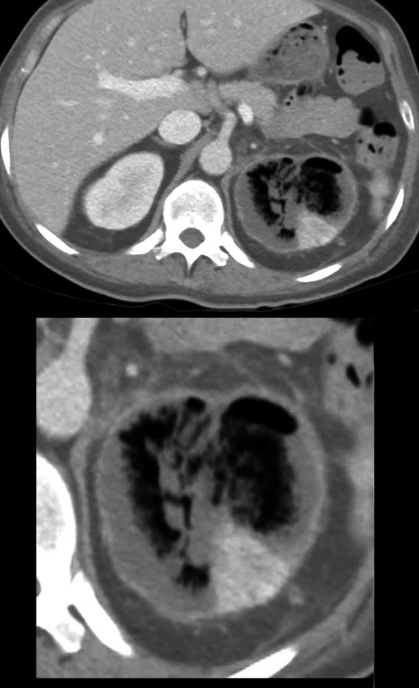
55-year-old diabetic female presents with flank pain and fever. CT with contrast in the nephrogram phase shows a non-functioning rim of cortex and medulla, and a small island of functioning parenchyma in the posterolateral portion of the left kidney. There is a thin enhancing rim that likely represents the enhancing capsule.
Ashley Davidoff MD TheCommonVein.net 135716c
- The patient was stabilized and discharged
- day 12 with
- continuous empiric CTX and Flagyl until her left kidney nephrectomy.
- procedure was delayed due to the COVID-19 pandemic.
- to 3 months after discharge)
- laparoscopic left nephrectomy.
- final diagnosis of
- xanthogranulomatous pyelonephritis from chronic pyelonephritis.
- surgical pus sample did not grow any organisms.
- She resumed work six weeks after her procedure.