

Selective angiography of the left renal artery in frontal projection (with images a and b magnified correspondingly in c and d) shows a cluster of vessels in the lower pole during the arterial phase. Shortly thereafter, an early draining vein is noted and the AVM becomes more homogeneous. These features are consistent with a renal AVM. It was embolized successfully.
Ashley Davidoff MD TheCommonVein.net 36307c
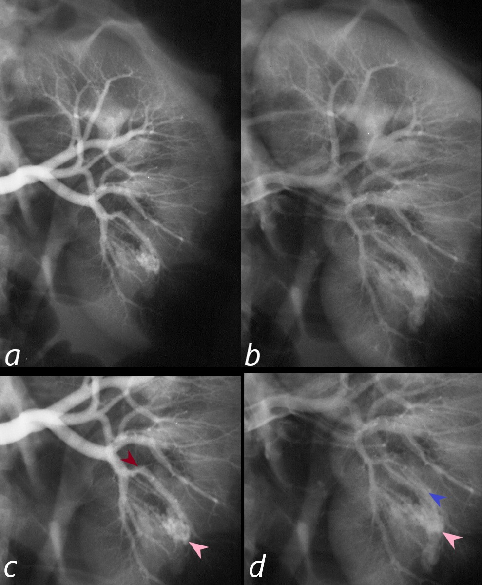
Selective angiography of the left renal artery in frontal projection (with images a and b magnified correspondingly in c and d) shows a cluster of vessels in the lower pole (c pink arrow) during the arterial phase (c maroon arrow is the feeding artery). Shortly thereafter (b magnified in d) an early draining vein is noted (d, blue arrowhead) and the AVM (d, pink arrowhead) becomes more homogeneous. These features are consistent with a renal AVM. It was embolized successfully.
Ashley Davidoff MD TheCommonVein.net 36307cL